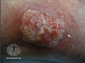
Cutaneous Squamous Cell Carcinoma
Cutaneous squamous cell carcinoma (cSCC) is a malignant skin tumor that arises from the epidermal keratinocytes. They can develop on any cutaneous surface, including the head, neck, trunk, extremities, oral mucosa, periungual skin, and anogenital areas. In individuals with fair-skin, it mainly arises in sun-exposed areas. On the other hand, in individuals with darker skin tones, it primarily develops in non-sun exposed areas, such as the lower legs, anogenital region, and areas of chronic inflammation or scarring. The latter accounts for 20-40% of cSCC in black patients.
More than 90% of cases of SCC are associated with countless DNA mutations in various somatic genes. For example, mutations in the p53 tumor suppressor gene are caused by exposure to ultraviolet radiation (UV), especially UVB (signature 7). Other signature mutations are related to cigarette smoking, aging, immunosuppression.
Clinical features:
Cutaneous SCC presents with a wide variety of cutaneous lesions, including papules, plaques, or nodules, that can be smooth, hyperkeratotic, firm/indurated (more with well-differentiated), or ulcerated, fleshy, granulomatous, +/- area of hemorrhage or necrosis (poorly differentiated). Cutaneous SCC in situ (Bowen’s disease) mainly presents as an erythematous, well-demarcated, scaly patch or plaque located in sun-exposed areas. Lesions of invasive cSCC are often asymptomatic but may be painful or pruritic or have local neurologic symptoms (e.g., numbness, stinging, burning, paresthesias, paralysis, or visual changes).
Diagnosis:
SCC is diagnosed based on clinical features and confirmed pathologically by diagnostic biopsy or following excision. Staging investigations may also be carried out in high-risk SCC to determine whether it has spread to lymph nodes or elsewhere.
Cutaneous SCC is catigorized as low-risk or high-risk, depending on the chance of tumor recurrence and metastasis. Characteristics of high-risk SCC are shown in this Figure. Metastatic SCC is found in regional lymph nodes (80%), lungs, liver, brain, bones, and skin.
Management:
Cutaneous SCC is almost always treated surgically, and most cases are excised with a 3–10 mm margin of normal tissue around the tumor. Reconstruction using a flap or skin graft may be needed to repair the defect. Other methods of removal include:
- Shave, curettage, and electrocautery: for low-risk tumors on trunk and limbs
- Aggressive cryotherapy: for tiny, thin, low-risk tumors
- Mohs micrographic surgery: for large facial tumors with indistinct margins or recurrent tumors
- Radiotherapy: for an inoperable tumor, patients unsuitable for surgery, or as adjuvant
For advanced/or metastatic SCC?
Locally advanced primary, recurrent or metastatic SCC requires multidisciplinary team (MDT) consultation. Usually, a combination of treatments is used, including surgery, radiotherapy, cemiplimab, and experimental targeted therapy using epidermal growth factor receptor (EGFR) inhibitors.
Written By: Dr. Iman Nazer, Dermatology resident.
References:
Uptodate
Dermnetnz.org
BAD guidelines


