Scarlet Fever
What is Scarlet Fever?
Scarlet fever is an infectious disease mediated by exotoxins, characterized by a distinctive erythematous rash affecting the skin and tongue. It typically arises from an infection of the throat, or less frequently, the skin, caused by Group A Streptococcus (GAS). Although scarlet fever can occur at any age, it predominantly affects children between the ages of 1 and 10 years, with the highest incidence observed in those aged 3 to 6 years. It is relatively rare in infants under 1 year of age and in adults.
Risk factors of Scarlet Fever
- Group A Streptococcus (GAS) Pharyngitis
- Presence in crowded environments
- People in close contact with someone who has a strep throat or skin infection
- Age range of 1 to 10 years
- Seasonal prevalence, particularly during winter and spring months
Causes of Scarlet Fever
Scarlet fever is caused by a bacterial infection Streptococcus group A (Streptococcus pyogenes), a type of bacteria that is commonly found in the throat and on the skin. This group of bacteria can produce toxins that trigger the symptoms of scarlet fever, including a red rash, fever, and a sore throat.
Clinical features of Scarlet Fever
Scarlet fever typically manifests with abrupt onset of fever, often accompanied by pharyngitis, cervical lymphadenopathy, headache, nausea, vomiting, anorexia, swollen and red strawberry tongue, abdominal pain, myalgia, and general malaise.
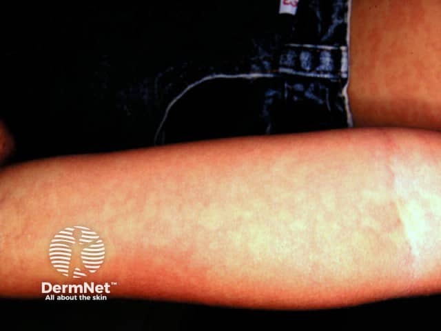
The characteristic erythematous rash usually emerges 12 to 48 hours following the onset of fever, initially affecting areas such as the neck, below the ear, chest, axillae, and groin, before progressively spreading to the remainder of the body within the subsequent 24 hours.
The initial rash in scarlet fever often manifests as erythematous spots or blotches, giving the skin a “boiled lobster” appearance. As the lesions expand and become more widespread, they may resemble sunburn, with goose pimples. The affected skin often develops a rough, sandpaper-like texture. In skin folds, particularly in the axillary and cubital areas, capillary rupture can lead to the formation of characteristic red streaks, known as Pastia’s lines, which may persist for 1 to 2 days after the generalized rash has resolved. A typical red, flushed appearance of the cheeks accompanied by pallor around the mouth.
In untreated patients, the fever typically reaches its peak by the second day and gradually returns to baseline over a period of 5 to 7 days. However, when appropriate antibiotic therapy is administered, the fever generally abates within 12 to 24 hours. By the sixth day of infection, the rash begins to to fade and peeling, resembling that of sunburned skin. The peeling is most prominent in areas such as the axillae, groin, and the tips of the fingers and/or toes, and it may persist for up to 6 weeks.
Diagnosis of Scarlet Fever
Scarlet fever is typically diagnosed based on clinical history and physical examination findings. Diagnostic confirmation is supported by throat swab culture or a rapid streptococcal antigen test, ideally obtained from the posterior pharynx or tonsils.
Additionally, elevated titers of anti-deoxyribonuclease B (anti-DNase B) and antistreptolysin O (ASO) may further support the diagnosis.
Treatment of Scarlet Fever
Upon confirmation of a streptococcal infection, a 10-day course of antibiotics, typically penicillin, is prescribed. In patients with a penicillin allergy, macrolides serve as an appropriate alternative antibiotic .In cases of recurrence caused by antibiotic resistance, cephalosporins may be used
Additional management strategies include:
- Administering paracetamol as needed to alleviate fever, headache.
- Encouraging the consumption of soft foods and adequate fluid intake, particularly cool liquids, in cases of severe throat pain.
- Utilizing oral antihistamines and emollients to alleviate pruritus associated with the rash.
Fever typically improves within 12-24 hours after starting antibiotics, and most patients recover within 4-5 days, although skin symptoms may take several weeks to fully resolve
Written by:
Atheer Alhuthaili, Medical Intern.
Revised by:
Naif Alshehri, Medical Intern
References:
DermNet.
Amboss
BMJ

