Mycosis Fungoides
Mycosis Fungoides (MF) is a form of non-Hodgkin lymphoma that is considered the most common type of primary cutaneous T-cell lymphoma (CTCL).MF is a neoplasia of malignant monoclonal T lymphocytes that generally invades the skin and causes cutaneous signs and symptoms.It is characterized clinically during early stages as erythematous scaly patches and plaques, or during advanced stages as tumors or erythroderma, with lymph node and/or visceral involvement.And histologically presents as an epidermotropic infiltrate of small-medium sized CD4+ T lymphocytes with cerebriform nuclei. Mycosis Fungoides (MF) is the most prevalent cutaneous T-cell lymphoma, accounting for 50-65% of cases. Typically seen in men [1.6-2:1] ratio and appears in late adulthood with 55-60 years as a median age of diagnosis. Even so, MF is a rare and uncommon condition, the incidence of MF in the United States is approximately 0.3-1.02 new cases per 100.000/year. MF follows an indolent clinical course over years, with an estimated 5-year survival rate of 87% and a median survival of 11.4 years. Also patients who present with involvement of lymph nodes or viscera have a median survival of <1.5%.
MF natural history is a classical slow progression from patches to plaques to tumors stage typically on unexposed areas such as the trunk, buttocks and thighs. Due to MF manifesting a variety of clinical and pathological presentations, atypical presentations of MF may be difficult to diagnose. Within this broad spectrum of clinical presentations, the World Health Organization (WHO) classified MF into 3 main variants or subtypes; folliculotropic MF, pagetoid reticulosis, and granulomatous slack skin.
Patch stage:
● Poorly defined, irregular, finely scaling, red patches of variable size often with atrophic (thin, wrinkled) skin
● Occurs mainly on sun-protected sites particularly the lower trunk, thighs and, in women, the breasts
● Often asymptomatic
● Hypopigmented variant: pale, finely scaly patches without atrophy in children and Fitzpatrick skin types
IV-VI
● Poikiloderma atrophicans vasculare is atrophic patch stage MF presenting with skin thinning, pigment
changes, and dilation of capillaries (telangiectasia)
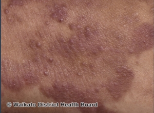
Plaque stage:
● Well-demarcated annular or arciform itchy thickened lesions
● Color often red, violaceous, or brown, sometimes scaly
Tumor stage:
● Large irregular lumps (>1 cm) developing from plaques
● Deep red to violaceous color, often shiny surface
Diagnosis:
The diagnosis of mycosis fungoides requires careful clinicopathological correlation. Dermoscopy can help to distinguish MF from inflammatory dermatosis.
Treatment:
Mycosis fungoides is treatable, not curable. There are no globally accepted treatment guidelines. Treatment should be guided by the patient’s wishes, stage of disease, availability of options, and local expertise.
Topical treatment:
● Topical steroids
● Topical chemotherapy eg, nitrogen mustard, carmustine
● Topical bexarotene
Procedural treatment:
● Phototherapy — PUVA, narrowband UVB
● Radiotherapy and whole-body total skin electron beam therapy
● Extracorporeal photopheresis
Systemic treatment:
● Chemotherapy — mycosis fungoides is relatively chemoresistant
● Immunotherapy — interferon-alpha
● Oral retinoids and rexinoids (eg, bexarotene)
Only brentuximab vedotin (for CD30+ MF) and allogeneic haematopoietic stem cell transplant alter the natural history of the disease.
Written by: Khalid Nagshabandi, medical student
Reference: DermNet