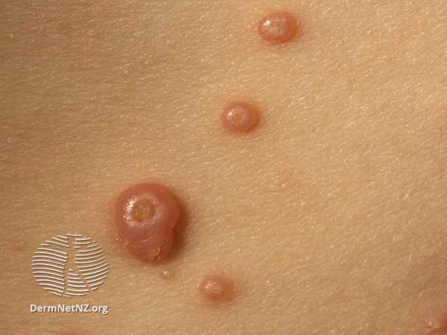
Molluscum Contagiosum
It is a benign, often asymptomatic viral infection of the skin, with no systemic manifestation. It is caused by molluscipoxvirus, a part of the poxviridae family which is a family of double-stranded DNA viruses. It is most common in childhood and early adolescence.
Risk Factors:
As mentioned, the etiology is due to molluscipoxvirus, but we have several risk factors associated with this disease. These risk factors include immunosuppression state, active atopic dermatitis, hot and humid climates, and crowded living conditions. Generally, those with poor immune function and immunocompromised skin are at risk for molluscum contagiosum.
Mode of Transmission:
Molluscum contagiosum can be transmitted due to direct contact during sports or other activities including sexual contact, indirect skin contact, or with fomites which are any object that can transmit the organism. Lastly, auto-inoculation can transmit the lesion by scratching or any trauma resulting in linear lesions (Pseudo koebner phenomenon).
Clinical Features:
The incubation period will usually be 2-6 weeks or more. Usually, the patient will come complaining of single or multiple non-tenders, skin-colored, pearly, dome-shaped papules with central umbilication. The lesion will be 2–5 mm in diameter and contain a caseous plug which if you squeeze will produce white cheesy fluid that consists of molluscum bodies.
The usual lesions in children will be the face, trunk, and limbs, in contrast to adults where the lesions will be usually in the abdomen, inner thighs, and genitalia. Finally, patients with HIV will usually have widespread and large (more than 15 mm) lesions.
Diagnosis:
Molluscum contagiosum is usually recognized by its characteristic clinical appearance (dome-shaped papules, often with central umbilication), and is distinctive on dermatoscopy which will show white molluscum bodies can often be expressed from the center of the papules. Sometimes, the diagnosis is made on skin biopsy. Histopathology will show the following features, acanthosis, cup-shaped invagination, and molluscum bodies (Henderson-Paterson bodies) which are keratinocytes with eosinophilic intracytoplasmic inclusion bodies containing viral particles.
Management:
Spontaneous remission is expected, however active treatment is considered in sexually transmitted molluscum, immunocompromised individuals or upon parental request.
The first line of treatments includes cryotherapy and curettage which has an immediate resolution of the lesion. Both treatments are well tolerated by adults.
Cantharidin which is a blistering agent can be applied directly to lesions, and a small blister will form which will result in the disappearance of the lesion and healing without scarring. Podophyllotoxin can also be used directly on the skin.
Immunocompromised patients or individuals with refractory disease will usually benefit from Cidofovir, Interferon-α, and Imiquimod. Other treatments for molluscum include potassium hydroxide, salicylic acid, topical retinoids, oral cimetidine, and pulsed dye lasers.
Complications:
It includes secondary bacterial infection from scratching, conjunctivitis, disseminated secondary eczema, and scarring.
Prevention:
Patients should be instructed to do the following, keep good hygiene, cover the lesion, do not share towels, clothing, or other personal items, and avoid sexual activity until the resolution of the lesion.
Written by:
Mohammed Alahmadi, Medical Student.
Revised by:
Maee Barakeh, Medical Student.
References:
DermNet
Dermatology Essentials by Bolognia
Fitzpatrick’s Color Atlas and Synopsis of Clinical Dermatology
UpToDate

