Dermatomyositis
Definition:
dermatomyositis is an autoimmune condition that often affects both the skin and the muscles (classic dermatomyositis), but it can alternatively manifest as skin-predominant illness (clinically amyopathic dermatomyositis). While children have no elevated risk of cancer, all adult patients presenting with cutaneous lesions of dermatomyositis should be investigated for concurrent muscle illness, systemic involvement, and/or malignancy.
Epidemiology:
Dermatomyositis exhibits a bimodal age distribution, affecting both adults ( 40-60 years) and children ( 4-14 years). It is a rare condition found worldwide. In adults, women are two to three times more likely to develop dermatomyositis than men. The disease occurs at a rate of 2 to 9 cases per million across different populations.
Etiology:
Dermatomyositis is thought to arise from an immune-mediated process triggered by external factors, such as malignancy, medications, or infections, in individuals with a genetic predisposition. Serum antinuclear autoantibodies are frequently detected, along with other myositis-specific autoantibodies. Antisynthetase antibodies target cytoplasmic antigens, meaning the antinuclear antibody test may sometimes be negative. Patients with antisynthetase antibodies often present with overlap syndromes. The term “antisynthetase syndrome” is used to describe individuals with these autoantibodies who exhibit symptoms like fever, erosive polyarthritis, “mechanic’s hands,” Raynaud’s phenomenon, and interstitial lung disease.
Clinical features:
Cutaneous manifestations:
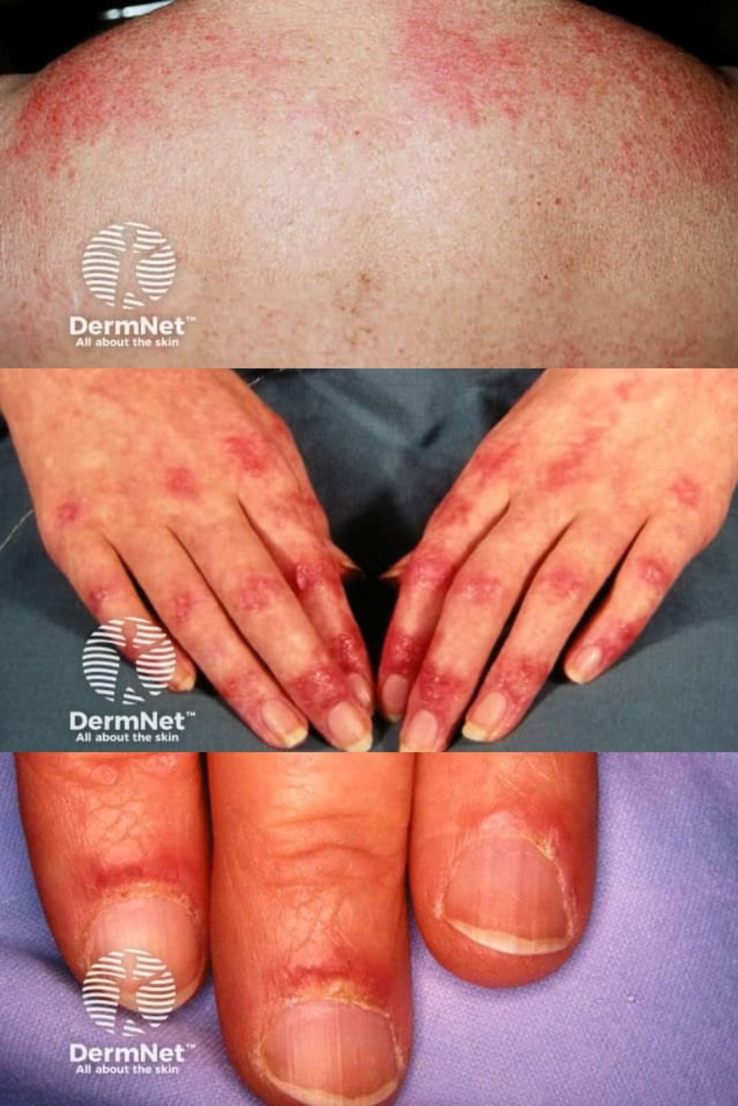
- The hallmark features of dermatomyositis include the heliotrope sign and Gottron papules. The heliotrope sign is identified by a pink-violet discoloration, mainly affecting the eyelids and periorbital skin, often accompanied by swelling. Dermatomyositis skin lesions are typically more prominent on extensor surfaces, such as the elbows, knees, metacarpophalangeal joints , and both proximal and distal interphalangeal joints (knuckles). When the papules on the knuckles develop a lichenoid texture, they are referred to as Gottron papules, while involvement of the elbows or knees is called Gottron sign.
- A key diagnostic feature of the dermatomyositis skin eruption is poikiloderma, which appears as a pink-violet color in dermatomyositis and a more reddish tone in lupus erythematosus. Photodistributed poikiloderma is highly characteristic of dermatomyositis, often seen on the upper chest (V-neck sign) and upper back (shawl sign). It can also occur in areas not exposed to sunlight, such as the lateral thigh, known as the holster sign. The skin lesions of dermatomyositis are frequently very itchy, which can significantly impact the patient’s quality of life. This itching can sometimes help distinguish dermatomyositis from lupus erythematosus.
- Calcinosis cutis is more common in juvenile dermatomyositis, affecting 25–70% of children. It appears as hard, irregular papules or nodules that may drain a chalky substance. These lesions tend to develop in areas of trauma, like the elbows and knees, but can occur elsewhere and may be painful, potentially interfering with function.
Systemic disease:
In dermatomyositis, skin symptoms often appear before noticeable muscle involvement; however, when muscle disease occurs, it is clinically similar to polymyositis. The myopathy primarily affects proximal muscles, particularly the extensor groups like the triceps and quadriceps, in a symmetrical pattern. In more severe cases, all muscle groups can be impacted. Patients may struggle with simple activities, such as combing their hair or standing up from a seated position.
Pulmonary complications occur in about 15–30% of dermatomyositis patients and typically present as diffuse interstitial fibrosis, resembling that found in individuals with rheumatoid arthritis or systemic sclerosis. Cardiac involvement is usually asymptomatic, but when it occurs, it typically manifests as arrhythmias or conduction abnormalities.
Diagnosis:
- A thorough history, including possible triggers and any past malignancies, along with a review of systems, should be conducted.
- Physical examination – This should include an assessment of the skin, muscles, and a complete general examination.
If the patient presents with a characteristic skin eruption, a skin biopsy is recommended to confirm the clinical diagnosis of dermatomyositis. Once histopathological confirmation is obtained, an evaluation for muscle and/or systemic disease should follow, as this will guide treatment decisions.
The assessment for myositis may involve testing the strength of proximal extensor muscles and other muscles, including the neck flexors, measuring serum muscle enzyme levels, performing an electromyogram, and/or conducting a triceps muscle biopsy. However, MRI or ultrasound of the proximal muscles is increasingly used instead of, or before, a muscle biopsy, especially when classic skin findings and consistent histology are present.
Additional baseline tests should be carried out to check for pulmonary, cardiac, or symptomatic esophageal diseases.
Management:
Skin-limited disease:
- First-line treatment: Photoprotection, topical corticosteroids (CS), and calcineurin inhibitors with or without antimalarials.
- Second-line treatment: High-dose methotrexate (MTX), mycophenolate mofetil (MMF), intravenous immunoglobulin (IVIG), cyclosporine, and rituximab, particularly for severe skin conditions with ulceration.
- Calcinosis cutis treatment: Diltiazem and possibly surgical removal; early aggressive treatment of juvenile dermatomyositis (JDM) to lower the risk of developing calcinosis cutis.
Disease monitoring:
-
- Muscle enzymes and clinical examinations should be rechecked every 2-3 months. If muscle disease develops, systemic steroids should be initiated.
- Physical exams every 4-6 months to screen for malignancy, especially during the first 2-3 years following diagnosis.
Skin and muscle disease:
- First-line treatment: Systemic corticosteroids (CS), methotrexate (MTX), and azathioprine (AZA).
- Second-line treatment: IVIG, mycophenolate mofetil (MMF), cyclosporine, cyclophosphamide, and rituximab.
Written by:
Mashael Alanazi, Medical Intern
Revised by:
Naif Alshehri, Medical Intern
Resources:
Bolognia textbook
Review of dermatology textbook

