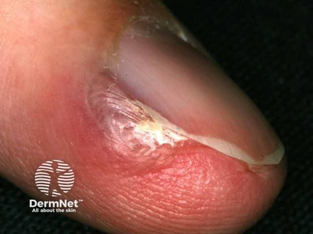
Glomus Tumor

Definition and Epidemiology:
A glomus tumour is a a rare, benign neoplasm composed of cells resembling the modified smooth muscle cells of the normal glomus body or the Sucquet-Hoyer canal. It is usually located in areas of the skin that are rich in glomus bodies (eg, the subungual regions of digits or the deep dermis of the palm, wrist, forearm, and foot) and can be extremely painful, particularly following change in temperature or pressure.
Solitary glomus tumors can occur at any age, but are most common in young adults. Although these tumors in general show no gender predilection, subungual lesions are more common in women.
Glomus tumors of the fingers and toes occur in approximately 5 percent of patients with neurofibromatosis type 1 (NF1) and are considered NF1-associated neoplasms.
Clinical features:
The glomus tumor is a benign lesion that usually presents in young adults (20–40 years of age) as a small (<2 cm), blue–red papule or nodule in the deep dermis or subcutis of the distal upper or lower extremities. They are tender to touch, and may be associated with severe paroxysmal pain in response to temperature changes and pressure. The hand, especially the nail beds and palm, is most commonly affected, but cutaneous lesions can also occur at other sites. Unusual extracutaneous glomus tumors have been reported in the gastrointestinal tract, bone, mediastinum, trachea, mesentery, cervix, and vagina. Extremely rare instances of malignant transformation within glomus tumors, with documented metastasis, have been described.
Diagnosis:
The diagnosis is suspected on the basis of the clinical appearance and history of paroxysmal pain and cold sensitivity. Histopathologic examination of the excised tumor is necessary to confirm the diagnosis.
Histologically, glomus tumor is a well-circumscribed dermal nodule composed of glomus cells, vasculature, and smooth muscle cells . Solid glomus tumor, with scarce vasculature and scant muscle component, is the most common variant. Less common variants include glomangioma, with prominent vascular component, and glomangiomyoma, with prominent vascular and smooth muscle components.
Treatment:
Treatment is surgical excision. For subungual tumors, preoperative imaging studies with color Doppler ultrasonography and magnetic resonance may provide information on tumor size, shape, and precise anatomic location.
Done by:
Bandar Alharbi, Medical Intern
Revised by:
Naif Alshehri, Medical Intern
Resources:
Dermnetz
UpToDate
Bolognia Textbook
