Pemphigoid Gestationis
Epidemiology:
Pemphigoid Gestationis is a rare, self-limited, autoimmune bullous disease. It is the most clearly characterized dermatosis of pregnancy and the only type of dermatosis of pregnancy that may also affect the skin of the newborn. The incidence of pemphigoid gestationis has been estimated at 1:20,000–1:50,000 pregnancies.
Pathogenesis:
Pemphigoid gestationis is caused by circulating immunoglobulin G1 (IgG1) autoantibodies directed against the 180 kilodalton bullous pemphigoid antigen (BP180 or collagen XVII), a transmembrane hemidesmosomal glycoprotein expressed in the basement membrane zone of the skin. As in bullous pemphigoid, the binding of antibodies to antigens within the basement membrane zone stimulates an inflammatory cascade that results in separation of the epidermis from the dermis. In pemphigoid gestationis, the primary site of autoimmunity seems to be the placenta, as antibodies bind not only to the basement membrane zone of the epidermis, but also to that of chorionic and amniotic epithelia, both of ectodermal origin.
Clinical features:
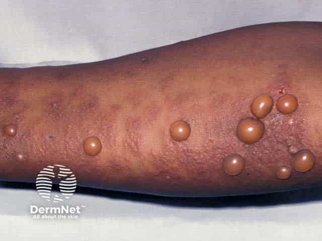
Pemphigoid gestationis most often present in the second or third trimester of pregnancy. Intense pruritus may precede the onset of visible skin lesions. The rash typically begins on the trunk as urticarial plaques or papules surrounding the umbilicus. Vesicles may also be present. Lesions may be seen on the palms and soles but rarely on the face or mucous membranes. The eruption spreads rapidly and forms tense blisters. The entire body surface may be involved, but the mucous membranes are usually spared.
Pemphigoid gestationis may remit prior to delivery. However, 75 percent of patients have a flare postpartum, and at least 25 percent of patients subsequently have a flare with use of oral contraceptive pills or during menses. Most cases spontaneously resolve in the weeks to months following delivery. The disease usually recurs with subsequent pregnancies and is often worse but may also skip pregnancies.
Diagnosis:
The diagnosis of pemphigoid gestationis is based upon the combination of clinical findings, examination of a lesional skin biopsy for routine histopathology and a perilesional skin biopsy for direct immunofluorescence (DIF). DIF reveals a homogeneous, linear deposit of complement C3 at the basement membrane zone. The presence of C3 is pathognomonic for pemphigoid gestationis in a pregnant patient.
Management:
The main goals of treatment of pemphigoid gestationis are to decrease blister formation, promote the healing of blisters and erosions, and relieve pruritus. In mild cases, the use of potent topical corticosteroids combined with emollients and systemic antihistamines may be adequate. However, systemic corticosteroids remain the cornerstone of therapy. Most patients respond to 0.5 mg/kg of prednisolone daily; the dose is tapered as soon as blister formation is suppressed. The common flare associated with delivery usually requires a temporary increase in dosage. Those rare patients with refractory disease may benefit from plasmapheresis during pregnancy.
Done by:
Bandar Alharbi, Medical Intern
Revised by:
Naif Alshehri, Medical Intern
Resources:
Dermnet
UpToDate
Bolognia

