Basal cell carcinoma (BCC)
Definition and Epidemiology:
Basal cell carcinoma (BCC) is a common skin cancer arising from the basal layer of epidermis and its appendages. These tumors have been referred to as “epitheliomas” because of their low metastatic potential. However, the term carcinoma is appropriate, since they are locally invasive, aggressive, and destructive of skin and the surrounding structures including bone. Estimates of the incidence of BCC are imprecise since there is no cancer registry that collects data on BCC. The American Cancer society estimates that in 2012, 5.4 million cases of nonmelanoma skin cancers (NMSCs) were diagnosed in 3.3 million people, of which approximately 8 in 10 cases would have been BCC.
Risk factors:
- BCC is particularly common in Caucasians; it is very uncommon in darker-skinned populations.
- The incidence in men is 30 percent higher than in women, particularly with the superficial type.
- The incidence of BCC increases with age; persons aged 55 to 75 have about a 100-fold higher incidence of BCC than those younger than 20.
- Exposure to UV light.
- Chronic arsenic exposure
- Radiation therapy
- Long term immunosuppressive therapy
- Basal cell nevus syndrome.
Clinical features:
Approximately 70 percent of BCCs occur on the face, consistent with the etiologic role of solar radiation. Fifteen percent present on the trunk, and only rarely is BCC diagnosed on areas like the penis, vulva, or perianal skin.
The clinical presentation of BCC can be divided into three groups, based upon lesion histopathology: nodular, superficial, and morpheaform.
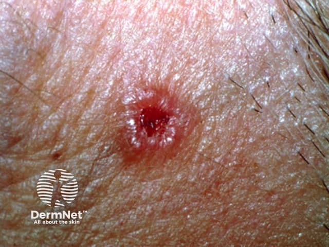
Nodular: Nodular BCCs, which represent about 60 percent of cases, typically present on the face as a pink or flesh-colored papule. The lesion usually has a pearly or translucent quality and a telangiectatic vessel is frequently seen within the papule. The papule may often be described as having a “rolled” border, where the periphery is more raised than the middle. Ulceration is frequent, and the term “rodent ulcer” refers to these ulcerated nodular BCCs.
Superficial: About 30 percent of BCCs are superficial BCCs. For unclear reasons, men have a higher incidence of superficial BCC than do women.
Superficial BCCs most commonly occur on the trunk, and typically present as slightly scaly, non-firm macules, patches, or thin plaques light red to pink in color. The center of the lesion sometimes exhibits an atrophic appearance and the periphery may be rimmed with fine translucent papules. A shiny quality may be evident when a superficial BCC is illuminated.
Morpheaform: Morpheaform or sclerosing BCCs constitute 5 to 10 percent of BCCs. These lesions are typically smooth, flesh-colored, or very lightly erythematous papules or plaques that are frequently atrophic; they usually have a firm or indurated quality with ill-defined borders.
Diagnosis:
Clinicians who are familiar with the clinical manifestations of BCC are often able to make the diagnosis based upon clinical examination. Even so, a skin biopsy is usually performed to provide histologic confirmation of the diagnosis.
Treatment:
The treatment for a BCC depends on its type, size and location, the number to be treated, patient factors, and the preference or expertise of the doctor. Most BCCs are treated surgically. Long-term follow-up is recommended to check for new lesions and recurrence; the latter may be unnecessary if histology has reported wide clear margins.
Treatment options include:
- Excision biopsy
- Mohs micrographically controlled surgery involves examining carefully marked excised tissue under the microscope, layer by layer, to ensure complete excision.
- Superficial skin surgery comprises shave, curettage, and electrocautery. It is a rapid technique using local anaesthesia and does not require sutures.
- Cryotherapy is the treatment of a superficial skin lesion by freezing it, usually with liquid nitrogen.
- Photodynamic therapy (PDT) refers to a technique in which BCC is treated with a photosensitising chemical, and exposed to light several hours later.
- Topical imiquimod
- Topical 5-Fluorouracil
- Radiotherapy
Written by:
Bandar Alharbi, Medical Intern.
Revised by:
Naif Alshehri, Medical Intern.
Resources:
UTD
Dermnet
Mayo clinic