Pyogenic Granuloma
Introduction
Pyogenic granuloma, also known as lobular capillary hemangioma, granuloma gravidarum, or pregnancy tumor, is a benign vascular lesion characterized by the rapid proliferation of capillary blood vessels in the skin or mucosa. Despite its name, it is neither pyogenic nor granulomatous. Modern research shows it arises due to reactive neovascularization without any infectious involvement.
Epidemiology
Pyogenic granulomas (PGs) can develop at any age but are most common in children, teenagers, and young adults. A slight male predominance is observed overall, except for oral lesions, which are more frequent in females due to hormonal influences like pregnancy or oral contraceptive use. Rare presentations include neonates with multifocal PGs or adults with eruptive disseminated forms.
Etiology and Pathogenesis
PGs are often linked to pre-existing trauma or irritation, hormonal changes, or specific medications. Minor trauma accounts for 7% of cases, while oral lesions are commonly associated with chronic irritation and poor dental hygiene. Hormonal changes during pregnancy or contraceptive use are significant factors, with 5% of pregnant women developing gingival PGs.
Medications like retinoids, protease inhibitors, and cancer-targeted therapies can also trigger PGs. Additionally, molecular studies have identified mutations in the BRAF and NRAS genes, which activate the MAPK signaling pathway, suggesting a neoplastic component in some cases.
Clinical Features
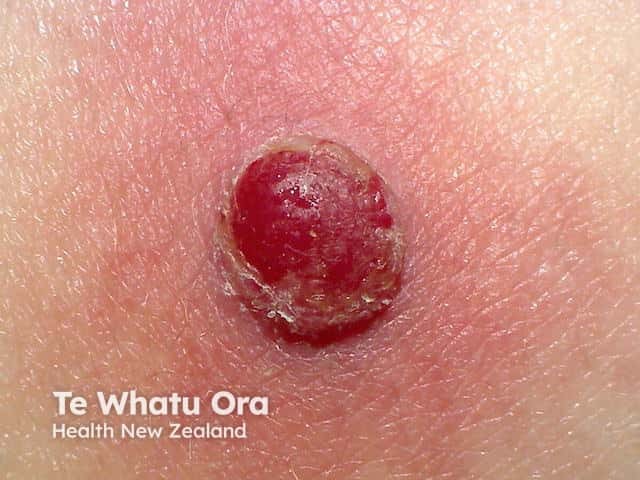
PGs typically present as painless, red nodules or polyps that grow rapidly over weeks. The lesions are highly friable, frequently ulcerating or bleeding after minor trauma. They stabilize at sizes usually under 1 cm but may persist indefinitely if untreated. Common sites include the gingiva, lips, fingers, face, and tongue.
Oral mucosal PGs often develop as pedunculated or sessile nodules on the gums or lips. These lesions may ulcerate, develop a yellow fibrinous surface, and cause minor discomfort. Rarely, satellite lesions may arise near a primary PG after trauma or irritation.
Dermoscopy and Histology
Dermoscopy
Dermoscopy reveals a distinct keratinized border forming a white collarette around the lesion. Vascular structures are prominent, with red homogeneous areas and white linear “rail lines” being hallmark features.
Histological Findings
Histologically, PGs are characterized by a lobular arrangement of capillaries in the dermis surrounded by inflammatory cells and fibromyxoid stroma. The epidermis is often thinned or ulcerated, and a peripheral collarette formed by elongated rete ridges is typically present. Signs of hemorrhage and inflammatory infiltrates, including lymphocytes and plasma cells, are common. Oral PGs may show an additional granulation tissue-like vascular proliferation pattern.
Differential Diagnosis
The differential diagnosis for PG depends on its location and presentation:
Cutaneous Differential Diagnosis
- Amelanotic melanoma: Most critical to exclude due to overlapping features.
- Bacillary angiomatosis: Especially in immunosuppressed patients.
- Kaposi sarcoma: Seen in HIV-positive individuals.
- Cherry angioma: Benign capillary proliferation.
- Irritated melanocytic nevus or wart: May mimic PG due to irritation.
Oral Differential Diagnosis
- Peripheral giant cell granuloma
- Peripheral ossifying fibroma
Histological Differential Diagnosis
- Cherry angioma
- Bacillary angiomatosis
Treatment
Treatment focuses on lesion removal and addressing contributing factors to prevent recurrence. Options include:
-
- General Measures: Improving oral hygiene, removing trauma sources, and discontinuing triggering medications.
- Topical Treatments: Timolol gel, imiquimod cream, cryotherapy, or table salt therapy (applied under occlusion).
- Procedural Treatments:
-
- Shave excision with electrosurgery: The most common approach.
- Curettage and cautery: Effective but with recurrence risk.
- Pulsed dye laser: Safe and effective for smaller lesions, particularly in children.
- Sclerotherapy: Using monoethanolamine oleate for minimal scarring.
- Surgical excision: Offers lower recurrence rates, especially in gingival lesions.
Prognosis
Pyogenic granulomas are benign and rarely resolve spontaneously, except in postpartum cases. Recurrence is common, particularly in gingival lesions or cases with persistent irritation or inadequate treatment. With appropriate intervention, the prognosis is excellent.
Written by:
Naif Alshehri, Medical Intern.
Resources:
Dermnet
Bolognia Textbook 5th Edition