Congenital Dermal Melanocytosis
Definition:
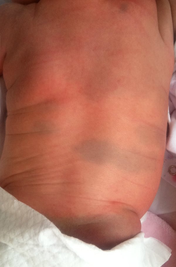
Congenital Dermal Melanocytosis, commonly known as Mongolian spots, is a congenital dermal pigmentation that appears as blue-grey patches on the skin, typically affecting the lower back and buttocks of newborns. It is a benign form of birthmark caused by the presence of melanocytes in the dermis.
Epidemiology:
Dermal melanocytosis is usually present at birth or develops within the first few weeks of life. In most cases, the lesions regress during early childhood, though persistence into adulthood has been documented, with a prevalence of 4% in Japanese males aged 18–22 years.
The incidence of dermal melanocytosis varies significantly based on ethnicity, occurring in nearly 100% of Malaysian, Mongolian, Japanese, Chinese, and Korean newborns; 87% of Bolivian Indigenous populations; 65% of Black individuals in Brazil; 17% of White individuals in Bolivia; and only 1.5% of White persons in Brazil. These variations suggest a genetic influence on the persistence of dermal melanocytes. Histologically, traces of dermal melanocytosis have been found in the presacral area of all newborns, regardless of race.
Etiology:
The characteristic blue-grey color of dermal melanocytosis results from the presence of melanin-producing melanocytes in the middle to lower dermis. During fetal development, melanocytes appear in the dermis around the 10th week of gestation and typically migrate to the epidermis or undergo apoptosis. However, in certain areas—such as the sacral region, scalp, and distal extremities—some melanocytes remain in the dermis, leading to persistent pigmentation.
The bluish appearance is due to the Tyndall effect, in which shorter wavelengths (blue and violet) are more likely to be scattered and reflected than longer wavelengths (red, orange, and yellow), giving the lesions their characteristic coloration.
Clinical Features:
Lumbosacral dermal melanocytosis typically presents as bluish-grey macular patches on the sacrococcygeal, lumbar, and gluteal regions. The patches can be round, oval, or irregular in shape, ranging in size from a few centimeters to over 20 cm. While they are usually limited to less than 5% of the body surface area, extrasacral variants can involve other areas such as the back, limbs, or face.
The affected skin remains unchanged apart from the pigmentation—there is no thickening, textural alteration, or other skin abnormalities. Persistent or extensive dermal melanocytosis may be associated with phakomatosis cesioflammea or phakomatosis cesiomarmorata, warranting further evaluation in such cases.
Diagnosis:
The diagnosis of dermal melanocytosis is clinical, based on the characteristic appearance and location of the lesions. No further testing is typically required unless an associated condition is suspected.
Management:
Dermal melanocytosis is a benign condition that usually fades spontaneously by the age of four. No treatment is necessary in most cases. However, in instances where lesions persist into adulthood, cosmetic camouflage or laser therapy (such as those used for nevus of Ota) may be considered for aesthetic reasons.
Written by:
Raneem Alahmadi, Medical Intern
Revised by:
Naif Alshehri, Medical Intern
Resources:
Bolognia 5th edition
Dermnet