Erythema Dyschromicum Perstans
Background
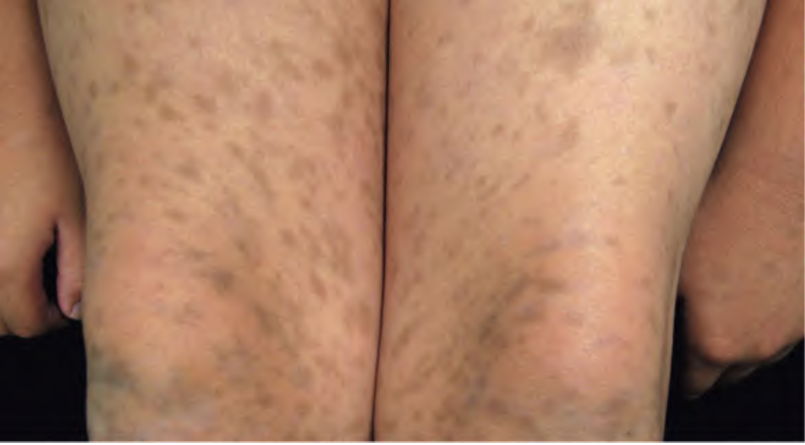
Ashy dermatosis, also known as erythema dyschromicum perstans (EDP), is a rare, chronic, idiopathic skin disorder characterized by the gradual appearance of bluish-gray to slate-colored macules and patches. It primarily affects individuals with darker skin phototypes (Fitzpatrick III–VI), particularly those from Latin America, Asia, and the Middle East. The condition is more commonly seen in young adults, but it can affect individuals of all ages. The term “ashy” refers to the gray color of the lesions, which gives the skin a dusky or dirty appearance.
Etiology
The exact cause of ashy dermatosis remains unknown, making it a diagnosis of exclusion. Several hypotheses have been proposed, including a delayed-type hypersensitivity reaction or cell-mediated immune response targeting basal keratinocytes. Histopathological findings often resemble a lichenoid tissue reaction, further supporting this theory.
There have been occasional associations with infections (e.g., hepatitis C), medications (e.g., proton pump inhibitors, antibiotics, and antiepileptics), and autoimmune conditions. However, these associations are not consistent and are often anecdotal.
Clinical Features
Ashy dermatosis typically begins with the insidious onset of slate-gray or bluish macules, which may be preceded by a faint erythematous border in early lesions. The patches are usually symmetrical, with a predilection for the trunk, neck, arms, and face. Lesions are non-scaly, non-itchy, and may gradually enlarge over time, coalescing into larger patches.The disease course is chronic and can persist for years.
Diagnosis
The diagnosis of ashy dermatosis is clinical, supported by histopathological examination when necessary to rule out mimickers. Skin biopsy typically shows interface dermatitis with basal cell vacuolization, melanin incontinence, and a perivascular lymphocytic infiltrate in the superficial dermis. However, these findings are not pathognomonic.
Dermoscopy may aid in identifying gray-blue dots and globules corresponding to dermal melanin. Differential diagnoses include lichen planus pigmentosus, post-inflammatory hyperpigmentation, macular amyloidosis, and drug-induced pigmentation. Careful clinical history and distribution of lesions help in differentiation.
Management
Treatment of ashy dermatosis is often challenging, as the condition tends to be resistant to therapy and cosmetically distressing for patients. There is no universally effective treatment, and management focuses on patient education, psychosocial support, and trial of medications with anecdotal or limited evidence.
Topical corticosteroids, calcineurin inhibitors (e.g., tacrolimus), and systemic agents such as clofazimine, dapsone, and isotretinoin have been tried with variable success. Some reports suggest that Q-switched lasers may provide pigment improvement, though recurrence is common. Sun protection is recommended as a general skincare measure, although sun exposure is not a known trigger.
Prognosis and Prevention
Ashy dermatosis is a benign condition without systemic involvement, but its chronicity and cosmetic impact can lead to psychological distress. Lesions may stabilize over time but often persist indefinitely. There is no established method for prevention, and recurrence after treatment is common.
Written by:
Dr Renad AlKanaan
Revised by:
Naif Alshehri, Medical Intern
References:
Dermnet
UpToDate
Bolognia 5th Edition
Picture Reference:
Bolognia 5th Edition