Acrodermatitis Enteropathica

Acrodermatitis enteropathica (AE) is a rare genetic disorder characterized by impaired zinc absorption, resulting in zinc deficiency. This condition affects the skin, gastrointestinal system, and immune system.


Pathophysiology:

Acrodermatitis enteropathica is primarily caused by mutations in the SLC39A4 gene, which encodes for the zinc transporter protein known as ZIP4. This protein plays a crucial role in facilitating the absorption of dietary zinc from the intestines into the bloodstream. Mutations in the SLC39A4 gene lead to impaired zinc uptake, resulting in zinc deficiency despite an adequate zinc intake.


Clinical Features:

Clinical manifestations usually appear within 1 to 2 weeks after weaning or at 4 to 10 weeks of age if bottle-fed.

Acrodermatitis enteropathica is characterized by a classic triad of symptoms, including periorificial dermatitis, diarrhea, and alopecia. 

  1. Periorificial Dermatitis:

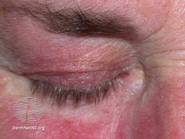
The rash in acrodermatitis enteropathica typically presents with a distinct distribution. The periorificial rash is commonly observed in a horse-shoe or U-shaped pattern, involving the areas around the mouth and eyes, while sparing the lips. There is usually a sharp demarcation between the affected area and normal skin, creating a noticeable contrast. The rash may manifest as eczematous or psoriasiform plaques that can progress to include vesicles, bullae, pustules, erosions, or hyperkeratotic areas.

  1. Diarrhea:

Diarrhea is a common symptom in acrodermatitis enteropathica and is often chronic in nature. 

  1. Alopecia:

Another characteristic feature of acrodermatitis enteropathica is diffuse hair loss, which affects the scalp, eyebrows, and eyelashes. The hair loss may be patchy or diffuse and is attributed to the disruption of hair follicle development and maintenance due to zinc deficiency.


In addition to the triad, acrodermatitis enteropathica can present with various additional cutaneous manifestations. These include glossitis, characterized by a red and glossy tongue, along with mouth ulcers and angular cheilitis. Symmetrical peri-anal excoriations and a rash on the buttocks can also occur. Zinc deficiency can impair wound healing, resulting in delayed healing of cuts and abrasions. Nail changes, such as softness, ridging, abnormal growth, and inflammation of the nail fold (paronychia), may also be observed.

While the noncutaneous symptoms of acrodermatitis enteropathica include blepharo-conjunctivitis, sensitivity to light, loss of appetite, irritability, depressed mood, growth failure in untreated children, hypogeusia, and hypogonadism in male adolescents and young adults.


Diagnosis:

The diagnosis of acrodermatitis enteropathica is primarily based on clinical features and confirmed by laboratory tests. A thorough physical examination, including a detailed evaluation of the skin and mucous membranes, can provide valuable diagnostic clues. Additionally, a detailed medical history, including feeding patterns and response to zinc supplementation, is essential.

Laboratory investigations play a crucial role in confirming the diagnosis. Blood tests can reveal low serum zinc levels, which is a strong indicator of zinc deficiency.


Management:

The management of acrodermatitis enteropathica involves lifelong zinc supplementation to correct the underlying deficiency. Zinc supplementation can be administered orally or, in severe cases, intravenously. The dosage and duration of zinc supplementation are individualized based on the severity of symptoms and response to treatment.

In addition to zinc supplementation, it is essential to address any associated complications. This may include the treatment of skin infections, nutritional support to correct deficiencies of other nutrients, and management of gastrointestinal symptoms such as diarrhea and vomiting.

In conclusion, acrodermatitis enteropathica is a rare genetic disorder characterized by impaired zinc absorption and resulting in zinc deficiency. It presents with a triad of symptoms involving the skin, gastrointestinal system, and immune system. Early diagnosis and lifelong zinc supplementation are crucial in the management of this condition to prevent complications and improve overall health outcomes for affected individuals.


Written by:

Deemah AlHuraish, Medical Student.

Revised by:

Maee Barakeh, Medical Student.


References:

DermNet.

NCBI.

Bolognia Textbook of Dermatology.

Benign Familial Pemphigus: Hailey–Hailey Disease

 Benign familial pemphigus, known as Hailey–Hailey disease, is an autoimmune uncommon hereditary blistering skin condition. This condition is unrelated to pemphigus vulgaris and has a different outcome.  Although it can happen at any age, benign familial pemphigus often manifests in the second to fourth decade. After that, it usually lasts a lifetime. All races are equally affected.

Etiology:

Although isolated occurrences of benign familial pemphigus without a family history can occasionally occur, the condition is hereditary usually. The ATP2C1 gene, which is located on chromosome 3q21–24, has now been confirmed to be defective. The calcium and manganese pump protein SPCA1 (Secretory Pathway Calcium/manganese-ATPase) is encoded by this gene. Desmosomes, which hold the keratinocytes together, appear to assemble improperly in the absence of enough calcium. The skin cells in benign familial pemphigus unstick from one another due to the genetic abnormality.

Clinical features:

In the skin folds, the disease often starts as a symmetrical, painful, erosive, and crusty skin rash. The armpits, groins, neck, under the breasts, and in between the buttocks are typical locations. The lesions usually fade away without leaving any scars. As the lesions get bigger, the center clears leaving a typical ring shape. If the lesions persist for a while, they could thicken. After that, the skin tends to macerate, producing painful cracks. These symptoms are exacerbated by heat, sweating, and friction. 

Diagnosis:

Benign familial pemphigus is often diagnosed based on appearance and family history, however, it is frequently confused with other skin conditions. Other blistering disorders, tinea cruris, thrush, and impetigo appear similarly. A skin biopsy may be needed for diagnosis. The histology is typical, with layers of detachment from the skin cells (acantholysis) lining up like a row of tombstones. The immunofluorescence test for antibodies comes out negative, in contrast to pemphigus vulgaris. Family members cannot yet access any diagnostic tests.

Management:

There is no definitive cure for this disease. Treatment is aimed at reducing symptoms and preventing flares. It is divided into topical and oral medication, in addition to general advice and other treatments. 

General advice includes avoiding triggers, washing, and drying skin folds carefully, and soft and loose clothing. Topical prescriptions include corticosteroids, antibiotics, benzoyl peroxide, ketoconazole, calcipotriol cream, and topical calcineurin inhibitors. Oral prescriptions include antibiotics, antivirals if herpes virus infection is a recurrent issue, and anticholinergic medications to reduce hyperhidrosis. In extreme circumstances, carbon dioxide laser vaporization or dermabrasion produces healing scars that are resistant to recurrence.

Complications: 

Secondary bacterial infection produces an unpleasant scent, also herpes simplex can cause blisters to form, and it has the potential to develop into a very painful viral illness (eczema herpeticum).

Prognosis: 

Many patients have long remissions, and an improvement with age does occur.

Written by:

Mohammed Alahmadi, medical student. 

Revised by:

Maee Barakeh, medical student.

References:

DermNet

Fitzpatrick’s Color Atlas and Synopsis of Clinical Dermatology

Review of Dermatology by Ali Alikhan

PAPA Syndrome

PAPA syndrome, also known as Pyogenic Arthritis, Pyoderma Gangrenosum, and Acne syndrome, is a rare autoinflammatory disorder characterized by a triad of symptoms: pyogenic arthritis, pyoderma gangrenosum, and severe acne. 

Pathophysiology:

The exact pathophysiology of PAPA syndrome is not yet fully understood. However, it is believed to be an autosomal dominant disorder caused by mutations in the PSTPIP1 gene, which is involved in regulating the immune system and inflammatory responses. The mutations lead to dysregulated production of pro-inflammatory cytokines, primarily interleukin-1 beta (IL-1β), resulting in chronic inflammation and tissue damage.

Clinical and Distinctive Features:

PAPA syndrome typically manifests in childhood or adolescence, although adult-onset cases have also been reported. The clinical features of PAPA syndrome include:

  1. Pyogenic Arthritis: Recurrent episodes of painful joint inflammation, particularly affecting large joints such as the knees, hips, and ankles. These episodes are characterized by swelling, warmth, and limited range of motion.
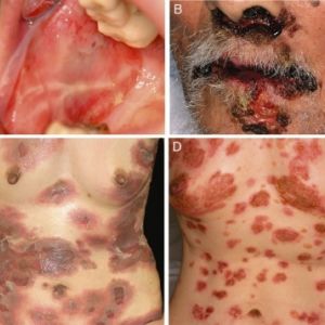
  1. Pyoderma Gangrenosum: Painful skin ulcers that develop in various areas of the body, most commonly on the lower extremities. These ulcers begin as pustules and progress to deep, necrotic wounds with undermined borders.
  1. Severe Acne: Persistent and severe acne, nodulocystic type, that is resistant to conventional treatments. It often involves the face, chest, and back, and can lead to scarring.

Diagnosis:

Diagnosing PAPA syndrome can be challenging due to its rarity and overlapping symptoms with other conditions. However, a comprehensive evaluation including a detailed medical history, physical examination, and laboratory tests can aid in the diagnosis. Key diagnostic criteria include the presence of the characteristic triad of symptoms and genetic testing to identify mutations in the PSTPIP1 gene.

Management:

The management of PAPA syndrome focuses on controlling symptoms, reducing inflammation, and preventing complications. Also, it involves a variety of approaches depending on the dominant manifestation:

Arthritis:

– Arthritis episodes often respond well to oral or intra-articular corticosteroids.

– In cases where corticosteroids are not satisfactory or arthritis recurs frequently, long-term corticosteroid use may be necessary, despite potential side effects.

Pyoderma Gangrenosum:

– Pyoderma gangrenosum may show some response to oral corticosteroids.

– Local immunosuppressant and anti-inflammatory drugs in the form of creams are commonly used.

– Treatment response is usually slow, but new biologic drugs targeting IL-1 or TNF have demonstrated efficacy in individual cases.

Acne:

– Acne treatment in PAPA syndrome may involve oral tetracycline antibiotics or isotretinoin, depending on the severity of the condition.

Developments in Treatment:

– Biological response modifiers, which target inflammatory proteins (cytokines), have shown success in managing other inflammatory conditions and are being explored for PAPA syndrome.

– Treatments targeting tumor necrosis factor (TNF) such as infliximab, etanercept, and adalimumab, as well as those targeting interleukin 1 (anakinra), have shown positive responses in cases of resistant arthritis and pyoderma gangrenosum.

Written by:

Deemah AlHuraish, medical student.

Revised by:

Maee Barakeh, medical student.

References:

DermNet

NCBI

Rosacea

Rosacea is a chronic skin disease that affects the central face and manifests with a variety of cutaneous or ocular symptoms.

Epidemiology:

Rosacea is primarily found among adults over 30 years old, and females are more likely to develop the disorder than males. People with light pigmentation or skin phototypes I and II are thought to be more prone to rosacea than those with darker pigmentation or skin phototypes III and IV.

Pathogenesis:

Rosacea has a poorly understood pathogenesis. Several potential contributing factors have been identified, including abnormalities in the innate immune system, inflammatory reactions to cutaneous microorganisms such as Demodex folliculorum and Staphylococcus epidermidis increased density on the skin , ultraviolet radiation exposure, vascular hyperreactivity, and genetics.

Clinical features:

Rosacea can be divided into four different types:

Type 1 – Erythematotelangiectatic Rosacea: Characterized by flushing and persistent central facial erythema with or without telangiectasia.

Type 2 – Papulopustular Rosacea: Characterized by persistent central facial erythema with transient papules and/or pustules.

Type 3 – Phymatous rosacea: Characterized by thickened skin with irregular surface and nodularities, particularly on the nose, chin or ears.

Type 4 – Ocular rosacea: Characterized by inflammations of the eyes and eyelids causing burning, stinging, dryness, and foreign body sensation of the eye.

Rosacea flares are usually associated with common triggers including sunlight, heat, stress, alcohol, and spicy foods.

Diagnosis:

For most patients, clinical assessment suffices to diagnose rosacea and rule out other disorders that might resemble it. The use of skin biopsies is rarely recommended, but can be beneficial if another disorder with specific histopathologic findings is suspected, or if granulomatous rosacea is suspected.

With highly pigmented skin, centrofacial erythema and telangiectasias can be subtle. A careful examination of other features of rosacea together with techniques such as dermoscopy and diascopy may help diagnose rosacea in patients with highly pigmented skin.

Management:

 Patient with rosacea should be instructed on the avoidance of triggers, gentle skin care routine including frequent moisturizing, avoiding exfoliant, and sunscreen use. 

Persistent erythema could be managed with brimonidine. Additionally, laser therapy or intense pulsed light can be used for facial erythema as well as facial telangiectasia.

Treatment with topical metronidazole, azelaic acid, or ivermectin is recommended for mild papules and pustules. Moderate to severe diseases or mild diseases that do not respond to topical agents should be treated with oral tetracycline, doxycycline, or minocycline. Treatment with oral isotretinoin may benefit patients with papules and pustules that are resistant to other therapies.

Rosacea can damage the ocular tissues when ocular involvement occurs. An ophthalmologist should be consulted if signs or symptoms of ocular involvement are present.

Written by:

Ghaida Altammami, medical student

Revised by:

Maee Barakeh, medical student

References:

UTD

DermNet

Bolognia textbook of dermatology

Fox-Fordyce Disease: The Sweat Retention Disease

Fox-Fordyce disease, also called apocrine miliaria, is a chronic rare condition where the apocrine sweat is retained and causes inflammation. Post-pubertal women between the ages of 13 and 35 are most commonly affected. Males and children can sometimes be affected. 

Etiology and pathophysiology:

There is no known cause of Fox-Fordyce disease. In the hair follicle, a keratotic block traps apocrine perspiration. The epidermis’s apocrine sweat ducts burst and leak, resulting in swelling and agonizing itching. There are several factors recognized for the development of this disease including alteration of sweat components, emotional and hormonal influences, and laser hair removal. Heat, humidity, friction, and stress are common environmental factors that contribute to the disorder.

Clinical features:

Apocrine sweat glands are exclusively located in the axilla, pubic region, and around the nipples, hence Fox-Fordyce disease is distinguished by itching lumps around the hair follicles in these places. The following clinical findings are often found with this disease includes bilateral dome-shaped flesh-coloured to small reddish papules affecting hair follicles, secondary skin lesion due to starching (lichenification), and reduced or absent sweating in the affected region. Loss of hair in the affected area is frequently observed. 

Diagnosis:

The diagnosis of Fox-Fordyce disease is typically made clinically; dermoscopy features include central hyperkeratotic plug in each papule, multiple light brown to dark structureless areas, lack of pigment network, and folliculocentric papule. 

Skin biopsy results show keratotic plug, spongiosis and vesicle formation, perifollicular infiltrate of chronic inflammatory cells, and xanthomatous foamy cells.

Management:

There is no definitive cure for this disease. One of the first treatments used is frequently topical and intralesional corticosteroids. Topical calcineurin inhibitors might lessen itching and make skin lesions look better. Topical tretinoin has been demonstrated to lessen pruritus but has little impact on how the condition manifests clinically and may irritate. Clindamycin lotion applied twice daily could also help to lessen symptoms. While some individuals have short relief from oral isotretinoin, some women respond to oral contraceptives.  Phototherapy, electrocautery, and periareolar skin excision are examples of physical therapies that could be helpful.

Prognosis: 

Fox-Fordyce disease could linger for a long time. It may occasionally go away during pregnancy. Others may experience a menopausal resolution or a continuing problem.


Written by:

Mohammed Alahmadi, medical Student. 

Revised by:

Maee Barakeh, medical student.

References:

DermNet

Bolognia Textbook of Dermatology

Fitzpatrick’s Color Atlas and Synopsis of Clinical Dermatology

Hidradenitis Suppurativa (HS)


Hidradenitis Suppurativa (HS), also known as acne inversa, is a chronic inflammatory skin condition that primarily affects specific areas of the body such as the axillae, groin, perianal, perineal, and inframammary locations. It is characterized by persistent or recurrent boil-like nodules and abscesses that culminate in a purulent discharge, sinuses, and scarring.


Etiology and Pathophysiology

Hidradenitis suppurativa (HS) is a complex skin condition with multiple causes. It is influenced by genetic, environmental, and behavioral factors. Genetic factors include a mutation in the gamma-secretase Notch signaling pathway found in some affected families. Environmental factors like obesity and smoking, as well as hormonal influences, contribute to the development of HS. The condition involves blockage of hair follicles, abnormal skin bacteria, inflammation, and damage to glands and ducts. The exact cause of HS is unknown, but it is likely a combination of genetic predisposition, hormonal changes, immune system dysfunction, and external factors that contribute to its development and severity.


Distinctive Features

HS typically manifests as painful nodules or boils in the affected areas, progressing to form abscesses and interconnected sinus tracts. These lesions have a tendency to recur and can persist for weeks or even months, resulting in the formation of scars and thickening of the skin. HS significantly impacts a person’s quality of life, causing persistent pain, discomfort, and emotional distress.
Clinically, HS is characterized by a range of specific features, including open double-headed comedones, painful firm papules and nodules, pustules, fluctuant pseudocysts, and abscesses. Additionally, the disease is marked by draining sinuses that link the inflammatory lesions, and it can lead to the development of hypertrophic and atrophic scars.

Diagnosis

Diagnosing HS can be challenging due to its variable presentation and similarities to other skin conditions. Clinical diagnosis requires recognition of the morphology (deep, inflamed, painful nodules, sinus tracts, scars), the location (intertriginous areas, apocrine gland-containing areas), and the chronicity of the disease process (prolonged course with periods of activity and remission).

The Hurley staging system is commonly used to classify the severity of HS:
Stage I: Single or multiple abscesses without sinus tracts or scarring.
Stage II: Recurrent abscesses with sinus tracts and scarring in two or more areas.
Stage III: Multiple interconnected sinus tracts and abscesses involving larger areas, with significant scarring.

In some cases, additional tests, such as swabs to rule out infection or biopsies for histopathological examination, may be performed to confirm the diagnosis or exclude other conditions.


Management:

Treatment options for hidradenitis suppurativa (HS) encompass various approaches based on the severity of the condition. These include:

  1. Lifestyle Modifications: Patients are advised to maintain good hygiene, avoid tight-fitting clothing, quit smoking, and manage weight if overweight or obese. These lifestyle modifications can help reduce friction, sweating, and inflammation in the affected areas.
  2. Topical Treatments: In milder cases, topical treatments such as antibacterial washes, topical antibiotics (e.g., clindamycin phosphate 1% with benzoyl peroxide), and anti-inflammatory creams can be effective. These interventions aim to reduce bacterial colonization and inflammation.
  3. Systemic Medications: For moderate to severe HS, systemic medications may be prescribed. Antibiotics like tetracycline or clindamycin can be administered to suppress bacterial growth. Anti-androgen medications like spironolactone may be considered for women with hormonal influences. Biologic therapies, such as TNF-alpha inhibitors like adalimumab, have shown promise in reducing inflammation and disease activity.
  4. Surgical Interventions: In cases where medical therapy proves insufficient, surgical interventions may be necessary. These can include incision and drainage of abscesses, excision of affected tissue, and laser therapy to manage persistent or recurrent lesions.


Written by

Deemah AlHuraish, medical student

Revised by

Maee Barakeh, medical student

References

DermNet

NCBI

Psoriasis: A Comprehensive Overview

Background

Psoriasis is a chronic inflammatory skin disease that is associated with a wide variety of comorbidities. It has also been considered a multisystem inflammatory disorder.

Epidemiology

It has been found that the prevalence of psoriasis among adults varied between 0.5 and 11.4 percent, while it ranged between 0 and 1.4 percent among children. The prevalence of psoriasis appears to be the same for males and females


Risk factors

Several factors are implicated in the development of psoriasis.

  • The genetic predisposition is considered to be one of the most important factors. Psoriasis susceptibility is linked to a number of genes. A major genetic determinant of psoriasis is the PSORS1 locus on chromosome 6p21 of the major histocompatibility complex (MHC).
  • The development of psoriasis can be exacerbated or caused by a variety of medical conditions, behavioral conditions, and environmental exposures. These include smoking, obesity, and alcohol consumption. Additionally, there are several drugs that can worsen psoriasis or psoriasis-like drug eruptions, such as beta blockers, lithium, and antimalarial drugs. It has been shown that infections, both bacterial and viral, can worsen psoriasis. In patients with psoriasis, low vitamin D levels have been observed, but their role in the disease remains uncertain. Psoriasis is often attributed to stress as an underlying or exacerbating factor.


Pathophysiology

An important role is played by T lymphocytes, dendritic cells, and cytokines (IL-23, IL-17, and tumor necrosis factor [TNF]) in psoriasis, which leads to hyperproliferation and abnormal differentiation of the epidermis, inflammatory infiltrates, and vascular dilation.


Clinical features

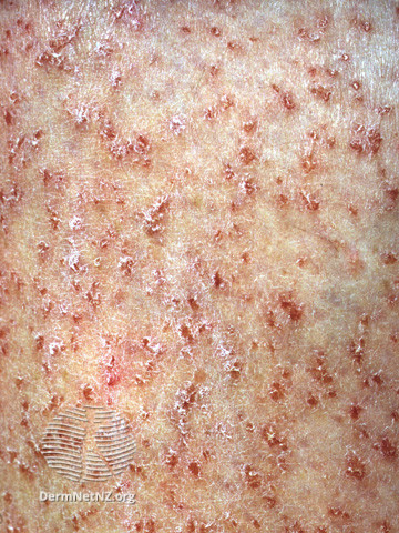
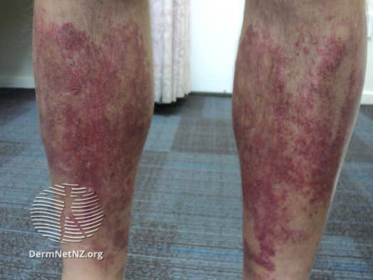
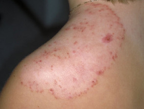
  • Chronic plaque psoriasis is the most common form of psoriasis, characterized by well-defined, erythematous plaques with an overlying, coarse scale. Commonly affected areas include the scalp, extensor elbows, knees, and gluteal cleft.
  • A guttate psoriasis usually manifests as a sudden eruption of numerous small, inflammatory plaques.  Affected sites are mainly the trunk and proximal extremities.
  • Pustular psoriasis can manifest as acute, subacute, or chronic pustular eruptions and has life-threatening complications. A severe form of generalized pustular psoriasis (von Zumbusch-type) presents with widespread erythema, scaling, and superficial pustules.
  • An erythrodermic is an uncommon type of psoriasis that is characterized by skin erythema that usually affects most or all of the body surface.
  • Inverse psoriasis, as the name implies, affects the intertriginous areas, including the inguinal, perineal, genital, intergluteal, axillary, and inframammary regions. The lesions are well-demarcated, smooth, shiny plaques with a minimal to nonexistent scale.
  • Nail psoriasis often develops after the appearance of psoriatic skin lesions. However, it can also occur concurrently or before psoriasis develops in other areas. The typical nail abnormality is pitting, which appears as a few to multiple tiny pits scattered over the nail plate.
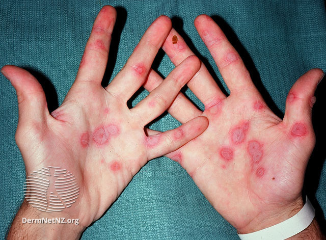
  • Psoriasis of the palms and soles is known as palmoplantar psoriasis. An erythematous, hyperkeratotic plaque with associated fissures is the classic presentation.

There are two classic signs of chronic plaque psoriasis: the Koebner phenomenon and the Auspitz sign. Koebner’s phenomenon describes the development of skin disease following skin trauma. Auspitz’s sign is characterized by pinpoint bleeding after the scaling overlying a plaque is removed.


Diagnosis

Physical examination is the most reliable method of diagnosing chronic plaque psoriasis in the vast majority of patients who suffer from the disease. It is sometimes necessary to perform a skin biopsy, which can be helpful in challenging cases, but it is not usually necessary.


Management

Psoriasis can be treated with a variety of topical and systemic treatments. It is important to consider the severity of the disease, relevant comorbidities, as well as the preferences of the patient when selecting treatment modalities

  • Limited plaque psoriasis:

Patients with limited plaque psoriasis should be treated initially with topical corticosteroids and emollients. Alternative treatments include tar, topical retinoids (tazarotene), topical vitamin D, and anthralin. In the case of facial or intertriginous areas, topical tacrolimus or pimecrolimus may be used.

  • Moderate to severe plaque psoriasis:

In most cases, it is advisable for most patients with moderate to severe plaque psoriasis to be initially treated with phototherapy.The use of systemic therapies may also be required. Commonly used medications include methotrexate and cyclosporine. Systemic corticosteroids are avoided due to the risk of severe withdrawal flare of psoriasis. In cases of severe psoriasis or psoriatic arthritis, biological therapy may be used. 


Written by

Ghida Altammami, medical Student

Revised by

Maee Barakeh, medical student

References

UTD

DermNet

Postinflammatory Hyperpigmentation (PIH)

Postinflammatory hyperpigmentation (PIH), also called acquired melanosis, is an acquired transient pigmentation following inflammatory conditions, skin damage, or therapeutic interventions. Darker skin types seem to experience it the most. PIH occurs with equal incidence in males and females of all ages.

Pathophysiology

Dermal melanosis occurs when inflammation disrupts the basal cell layer, causing melanin pigment to be released and subsequently trapped by macrophages in the papillary dermis. On the other hand, arachidonic acid is released during the epidermal inflammatory response, where it is oxidized to produce prostaglandins, leukotrienes, and other substances. These inflammatory byproducts affect melanocyte and immune cell function. These inflammatory substances specifically activate epidermal melanocytes, leading them to enhance melanin synthesis and subsequently increase pigment transfer to neighboring keratinocytes.

Causes

When PIH causes inflammation in the epidermis, melanocytes are stimulated to produce more melanin and transfer it to nearby keratinocytes. Acne excoriée, lichen planus, systemic lupus erythematosus, chronic dermatitis, and cutaneous T-cell lymphoma, particularly erythrodermic variations, are examples of common inflammatory illnesses that lead to PIH. A few drugs may also contribute to PIH. These include tetracycline, chemotherapeutic treatments like bleomycin, 5-fluorouracil, and busulfan, as well as antimalarial medications.

Clinical features

PIH presents at the original insult site after it has healed. The lesions’ coloring ranges from pale brown to black. If the patches are exposed to sunlight, they could get darker.

Diagnosis

The diagnosis of PIH is typically made clinically; no particular testing is necessary. Dermal PIH and epidermal PIH can be distinguished using a Wood lamp examination. To rule out additional underlying causes of hyperpigmentation, a skin biopsy is necessary. Melanin can be localized in the epidermis and/or dermis by staining the biopsy samples with Fontana-Masson silver stain.

Management

Treatment for PIH is frequently a challenging and lengthy process that frequently takes 6 to 12 months to produce the necessary depigmentation. Topical treatment for PIH aims to bleach the lesion and includes hydroquinone, azelaic acid, vitamin C cream, tretinoin, corticosteroids, glycolic acid, kojic acid, arbutin, and licorice extracts. Physical treatment for PIH includes chemical peels, laser treatments, and intense pulsed light therapies. If pigmentation affects an exposed site, daily application of broad-spectrum SPF sunscreen is important.

Prognosis

Without therapy, the clinical symptoms of epidermal melanosis could take months or even years to go away. People with dark complexion may experience PIH for a longer period of time than people with light skin. UV exposure and ongoing or recurrent inflammation can exacerbate the condition.

Written by

Mohammed Alahmadi, medical Student

Revised by

Maee Barakeh, medical student

References

DermNet

Medscape

UpToDate

Melasma

Background 

Melasma is a skin pigmentation disorder, characterized by hyperpigmentation of the skin due to hyperfunctioning melanocytes. It affects the sun-exposed areas of the face, such as the centrofacial, malar, and mandibular regions. It is most prevalent among young to middle-aged women.

Etiology

According to some theories, melasma can be attributed to UV irradiation exposure as the melanocytes of involved skin will be hyperfunctional and will release larger amounts of melanin pigment compared to uninvolved skin. Another theory suggests that melasma may be caused by skin photoaging that affects genetically predisposed individuals.

Risk factors 

There are multiple factors that increase the risk of developing melasma. These factors include genetics, exposure to sunlight, skin type, and hormonal factors (including pregnancy, hormonal therapies, and oral contraceptives).

Clinical features

Melasma presents with light to dark brown or brown-grey patches with irregular borders which appear primarily on the face. Precisely, it affects the forehead, nose, cheeks, upper lip area, and chin in a symmetrical pattern

There are three common facial patterns of melasma distribution: 

1- Centrofacial melasma generally affects the forehead, cheeks, nose, upper lip, and chin

2- Malar melasma mainly affects the cheeks

3- Mandibular melasma affects the lower jawline. 

Extra facial melasma, which is less common than the facial melasma and usually difficult to treat, can occur on the arms, forearms, chest, or back of some patients.

Diagnosis 

Melasma is typically diagnosed clinically. The Wood’s lamp examination can be helpful in identifying the location of pigment (epidermal or dermal), especially in those with lighter complexions. Dermoscopy has become increasingly useful in diagnosing melasma and determining its level of pigment deposition. 

Management 

All patients with melasma should use a broad-spectrum sunscreen with an SPF of 50 or higher on a daily basis.

The first-line treatment regimen for mild melasma is hydroquinone 4% cream. Patients with mild to moderate melasma use fluocinolone, hydroquinone, and tretinoin triple combination creams (TCC), rather than hydroquinone 4% cream alone.

Chemical peels (glycolic acid, other alpha-hydroxy acids, salicylic acid, Jessner’s peel, and trichloroacetic acid) are other treatment options for patients with melasma who have failed to respond to topical skin lightening therapies. 

For patients with melasma who have not responded well to topical treatments and chemical peels, lasers and light therapies can be provided.

Written by

Ghida Altammami, medical student

Revised by

Maee Barakeh, medical student

References

Bolognia textbook of dermatology

DermNet

UTD

Albinism

Albinism is a genetic disorder characterized by the partial or complete absence of pigment (melanin) in the skin, hair, and eyes. It results from a group of inherited genetic mutations that affect the production or distribution of melanin.


Etiology and Pathophysiology

Albinism is primarily inherited in an autosomal recessive manner, meaning that both parents must carry a mutated gene for their child to be affected. These genes are involved in the production of melanin or the enzymes required for its synthesis. The specific genes involved vary depending on the type of albinism. The most common types include oculocutaneous albinism (OCA) and ocular albinism (OA).

In OCA, the melanin production is impaired in the skin, hair, and eyes. Mutations in genes such as TYR, OCA2, TYRP1, and SLC45A2 are commonly associated with different types of OCA. In contrast, OA primarily affects the eyes, and mutations in the GPR143 gene are usually responsible.


Distinctive Features and Clinical Presentation

The hallmark dermatological feature of albinism is the absence or reduction of melanin pigment in the skin. This results in a strikingly light complexion, characterized by a pale or milky-white appearance. The skin is highly susceptible to sunburn and damage from ultraviolet (UV) radiation due to the lack of protective melanin. Therefore, individuals with albinism must take extra precautions to protect their skin from sun exposure.

The lack of melanin in the eyes can result in various eye abnormalities, such as nystagmus (involuntary eye movements), strabismus (misalignment of the eyes), and photophobia (sensitivity to light). Reduced pigmentation in the iris may cause the irises to appear light blue, green, or gray.

In addition to photosensitivity, individuals with albinism may experience other dermatological manifestations. These can include freckle-like macules, which are small, well-defined, light brown spots that may appear on sun-exposed areas over time. The macules are not true freckles but rather an accumulation of melanin precursor molecules due to impaired melanin production.


Diagnosis

The diagnosis of albinism is typically made based on clinical examination, family history, and specialized tests to assess visual function. Genetic testing may be performed to confirm the specific type of albinism and identify the responsible gene mutations.


Management

Management of albinism primarily focuses on minimizing the complications associated with the condition. One of the significant complications associated with albinism is an increased risk of developing skin cancers, including melanoma. The absence of melanin, which acts as a natural protective barrier against UV radiation, makes the skin more vulnerable to the harmful effects of the sun. Therefore, individuals with albinism must adopt strict sun protection measures to minimize the risk of skin cancers. These measures include using broad-spectrum sunscreen with a high sun protection factor (SPF), wearing protective clothing that covers the skin, seeking shade during peak sun hours, and regularly examining the skin for any suspicious lesions.

Regular dermatological examinations are essential for early detection of skin cancers in individuals with albinism. Dermatologists can perform thorough skin examinations and educate patients about self-examination techniques. Any suspicious skin lesions should be promptly evaluated and, if necessary, biopsied for further assessment

Visual aids, such as prescription glasses, contact lenses, or low-vision aids, can help improve visual acuity and alleviate the symptoms of nystagmus and strabismus. Regular eye exams are crucial for monitoring and managing any ophthalmic complications.

Psychological support and counseling are essential for individuals with albinism, especially during childhood and adolescence, as they may experience social and psychological challenges due to their appearance and visual impairments. Educating family members, peers, and educators about albinism can foster understanding and support.


Written by:

Deemah AlHuraish, Medical Student.

References:

DermNet

Medscape

Vitiligo: A Comprehensive Overview


What is Vitiligo

Vitiligo is a chronic, acquired, depigmenting, autoimmune disorder of the skin, resulting in the progressive loss of pigment-producing cells (melanocytes) which affect the color of the skin and hair. It manifests as milky-white patches of the skin, especially in those with dark skin, and it can be quite disabling from a cosmetic standpoint.

Epidemiology of Vitiligo

0.5–2% of people in the population have vitiligo. Between 20 and 24 years old is the typical onset age. Both sexes are equally affected, and there is a constant incidence across all races.

Causes of Vitiligo

Vitiligo is a multifactorial disorder related to both genetic and non-genetic factors. While environmental factors account only for 20% of vitiligo risk, genetic factors seem to account for 80% of it. The pathogenic hypotheses for vitiligo are variable and complex. The autoimmune destruction of melanocytes hypothesis suggests that autoantibodies are directed against several melanocyte antigens such as tyrosinase and tyrosinase-related proteins 1 and 2 (TYRP1 and TYRP2), which are crucial enzyme in the synthesis of melanin.

Clinical Features of Vitiligo

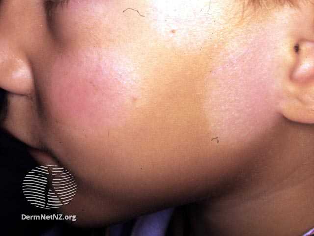
In most cases, vitiligo develops slowly. The most typical manifestation is a total loss of pigment in one or more macules or patches of the skin, which are characterized by their unique milky white color. Also, it typically has well-defined convex borders with possible hyper- or hypopigmented, red, or unaffected skin color borders. Vitiligo can be found in any region of the body; however, it tends to be more prevalent in sites of specific trauma, such as a cut, burn or abrasion, which is known as the Koebner phenomenon.

Classification of Vitiligo

Vitiligo classification is confusing. The following classification divides vitiligo into four general types. First, non-segmental vitiligo which tends to be bilateral and symmetrical. Second, segmental vitiligo which is usually unilateral and does not cross the midline. Third, mixed vitiligo, and lastly, unclassified vitiligo. These four classes are further divided into many subtypes. 

Diagnosis of Vitiligo

The diagnosis of vitiligo is typically made clinically; no particular testing is necessary. However, there are numerous instruments, such as the wood’s lamp, dermoscopy, and skin biopsy, that assist in the diagnosis.

Management of Vitiligo

There is no cure for vitiligo. The aim of treatment is to stop the progression of the disease and to achieve a satisfactory repigmentation. Minimizing skin injury and sun protection are important measures to limit disease progression. Topical treatments are commonly used which include corticosteroids, calcineurin inhibitors, and in some cases, JAK inhibitors cream. Phototherapy is also widely used, options include narrowband UVB, psoralen photochemotherapy(PUVA) and excimer laser which is useful in localized lesions of vitiligo. Systemic treatment may be needed, including systemic steroids, methotrexate, ciclosporin, and mycophenolate mofetil. Additionally, depigmentation therapy may be used in widespread disease with only a few areas of normally pigmented skin. 

Outcome of Vitiligo

There are some predictors that suggest an active disease, including peripheral hypopigmentation, confetti-like depigmentation, ill rather than well-defined borders, and the Koebner phenomenon. 

Vitiligo’s clinical course is incredibly variable. Vitiligo typically grows slowly over several months, then goes dormant for years. It can be challenging to manage.


Written by

Mohammed Alahmadi, medical student. 

Revised by

Maee Barakeh, medical student

References

Medscape

DermNet

Bolognia textbook of dermatology


Trichotillomania

Background

Trichotillomania is a hair pulling disorder, manifested by an uncontrollable desire to pull out one’s hair resulting in hair loss. It is often associated with skin picking. According to estimates, trichotillomania affects around 4% of the population, and it is more common in females with a 9:1 ratio. The onset of this disorder is usually at its peak during early adolescence and during the pre-school years.

Etiology & Pathophysiology

The cause is still uncertain, but some genetic factors have been identified as possible contributors. The pathogenesis of this disease could be linked to structural abnormalities or brain’s neurotransmitter imbalance. Moreover, it has been found that trichotillomania is largely associated with obsessive-compulsive disorder. In addition, it is crucial to be aware that stress and anxiety can also contribute to trichotillomania as a coping mechanism for the individual.

Clinical features 

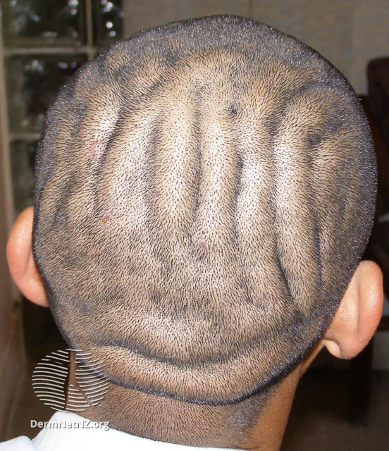
Various body sites can be affected by trichotillomania, including the scalp, face, arms, legs, and pubic region. The scalp, however, is the most common area for both children and adults. Due to the frequent pulling of hair at different times and at different sites over time, there is a noticeable variance in the length of the hair shafts. An individual’s pattern of hair loss can range from slightly thinning of the hair or unnoticeable hair loss patches to complete baldness.

Diagnosis

The diagnosis of trichotillomania is based upon history and physical examination.

According to the DSM-5, trichotillomania can be diagnosed as a disorder based on the following diagnostic criteria:

  • Hair loss caused by repeated hair pulling
  • Consistent attempts to stop or reduce hair pulling
  • At least one important area of functioning is significantly impaired by hair pulling
  • Hair pulls or hair loss are not associated with any other medical conditions
  • Hair pulling does not appear to be explained by another mental illness

It is essential to exclude other causes of hair loss before making a diagnosis. A skin biopsy can be performed in order to confirm the diagnosis and exclude any other causes of hair loss.

Management

The treatment should begin with reassurance and education of the parents and caregiver as well as the child. Patients are typically required to be seen by several different specialties, including primary care physicians, dermatologists, psychiatrists, and psychologists. Cognitive behavioral therapy (CBT) and habit reversal training have been found to be the most effective treatments for trichotillomania. Medications such as tricyclic antidepressants and selective serotonin reuptake inhibitors (SSRIs) may also be prescribed in resistant cases in adolescents and adults to manage symptoms.

Prognosis

Trichotillomania is typically benign and self-limited in children. An early diagnosis and treatment of the disorder improve the prognosis. In adolescents and adults, however, trichotillomania may be episodic, but tends to be chronic, making treatment more challenging.

Complications

There is a possibility that permanent hair loss may be one of the long-term complications of this disease. Moreover, an unusual but seriously dangerous complication of trichotillomania known as trichobezoar occurs when patients ingest pulled hair. There are also other complications that have been reported, such as skin infections, and blepharitis.

Written by

Ghida Altammami, medical student

Revised by

Maee Barakeh, medical student

References

National Library of Medicine

UpToDate

DermNet