Sunscreen Awareness Month
Introduction:
According to the American Academy of Dermatology Association, May is Skin Cancer Awareness Month, and May 28 is National Sunscreen Day, by the early 20-century sunscreen started to manufacture and by the mid-1970s that sunscreens gained a degree of consumer acceptance, which help and prevent a variety of dermatological conditions.
How to have optimal sun protection
To have good sunscreen:
Broad-spectrum sunscreen= UVA + UVB protection
Water resistance = maintenance of SPF after 40 or 80 minutes in water.
SPF ≥ 15 or 30 recommended.
Re-apply1-2 hours during outdoor activates
Other measures:
Staying in the shade especially from 10 AM – 2 PM
Wide brim hats
Sunglasses
Protective clothing
| Types of sunscreens: Chemical | Physical |
| works like a sponge on the surface of your skin, absorbing the sun’s rays. | works like a shield on the surface of your skin, deflecting the sun’s rays. |
| Optimal if you have sensitive skin | Easier to rub in the skin without leaving residue |
| Example: zinc oxide, titanium dioxide | oxybenzone, avobenzone, and ecamsule |
Concentrations as are utilized in SPF testing is: 2 mg/cm2 to achieve 2 mg/cm2 of density:
1 teaspoon of sunscreen to the face/head/neck
1 teaspoon to each upper extremity
2 teaspoons to the front and back torso
2 teaspoons to each lower extremity
Benefits of sunscreen:
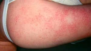
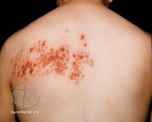
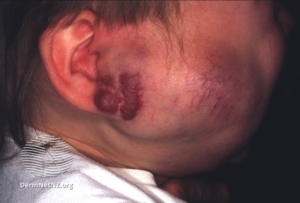
Reduce the onset of Photoaging
Photo immune Suppression
Non-melanoma Skin cancer
Photosensitivity Disorders
Written by: Abdullah Nasser Al-Omair, Dermatology Resident
Reference: UpToDate