Stasis Dermatitis (Venous Eczema)

Definition:

Venous dermatitis is a common type of eczema/dermatitis that involves one or both lower legs in association with venous insufficiency. It is also known as gravitational dermatitis.

Epidemiology:

Venous eczema primarily occurs in middle-aged and elderly individuals, with a prevalence of 20% in those over 70 years old. It is related with:

  • Stasis ulcers.
  • History of deep venous thrombosis in an affected limb
  • History of cellulitis 
  • Chronic edema in the lower leg, exacerbated by high temperatures and long periods of standing
  • Varicose veins

Etiology:

Venous dermatitis is caused by fluid accumulation in the tissues and triggering of the innate immune response.

Typically, during walking, the leg muscles facilitate the upward flow of blood and valves in the veins prevent blood from accumulating. Deep venous thrombosis (DVT) or varicose veins may compromise the valves in the deep leg veins. Back pressure causes fluid accumulation in the tissues. An inflammatory reaction occurs.

Clinical manifestation:

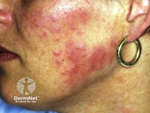
The condition usually manifests as red, scaly, and eczematous patches or plaques on chronically edematous leg. The medial ankle is most commonly and severely affected, with skin changes possibly extending to the knee and foot. Pruritus can lead to lichenification due to continuous scratching or rubbing.

Chronic forms are characterized by scaling and hyperpigmentation from cutaneous hemosiderin accumulation. Additional signs of chronic venous insufficiency and associated comorbidities are frequently observed, such as:

  • Atrophie blanche —refers to stellate, porcelain-white scarring patches caused by microthromboses. 
  • The lower leg shaped like “Champagne bottle” — narrowing at the ankles and induration (lipodermatosclerosis)
  • Ulceration

Complications:

  • Impetiginisation — is a secondary infection caused by Staphylococcus aureus that leads to the formation of yellowish crusts.
  • Cellulitis — an infection caused by Streptococcus pyogenes, characterized by redness, swelling, pain, fever, a red streak up the leg, and swollen lymph nodes in the groin.
  • Secondary eczema — spread of eczema to other parts of the body.
  • allergic reaction to one or more ingredients in the lotions or ointments used

Diagnosis:

Stasis dermatitis is usually diagnosed clinically on the basis of:

  • Skin lesions’ clinical presentation 
  • Venous insufficiency history
  • Other chronic venous insufficiency manifestations (e.g., varicose veins, pitting edema, hyperpigmentation) 

A skin biopsy is usually unnecessary to diagnose stasis dermatitis. If the diagnosis is unclear or if the patient presents with unusual characteristics (such as a single lesion or no ankle involvement), performing a biopsy and histopathologic examination can help confirm the diagnosis and rule out other skin conditions like allergic contact dermatitis, asteatotic eczema, or cutaneous malignancies. 

Management:

General measures:

  • Lifestyle modification— Patients with chronic venous disease and stasis dermatitis should make lifestyle adjustments to lower venous hypertension, decrease edema, and prevent venous ulcers. These consist of weight loss, regular walks, avoiding extended standing, exercising, and elevating your legs whenever possible. 
  • Skin care — To treat skin dryness and itching in mild or chronic stasis dermatitis, it is recommended to use mild, fragrance-free liquid cleansers for gentle skin washing and apply bland emollients regularly. White petroleum jelly is effective, affordable, and non-irritating. Emollients can be administered frequently throughout the day.

Compression therapy: 

Compression therapy using either compression bandaging systems or compression stockings, is the primary conservative treatment for stasis dermatitis and the associated chronic venous disease.


Written by:

Mashael Alanazi, Medical Student.

Revised by:

Maee Barakeh, Medical Student.

Resources:

DermNet.

Uptodate.

Pityriasis Alba

Pityriasis alba is a skin condition that primarily affects people between the ages of three and sixteen in children and young adults. It is common worldwide with a prevalence in children of around 5%. It affects males and girls female equally. Pityriasis alba hypopigmentation is more noticeable and possibly more prevalent in black skin types than in white skin types.

Etiology: 

The etiology of pityriasis alba is idiopathic. It is usually associated with atopic dermatitis and dry skin. It frequently appears after sun exposure, maybe because the afflicted spots become more noticeable due to surrounding skin darkening. Ongoing research about the relationship between pityriasis alba and the following factors: UV radiation, insufficient or excessive bathing, low levels of copper, and Malassezia yeasts.

Clinical features:

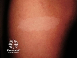
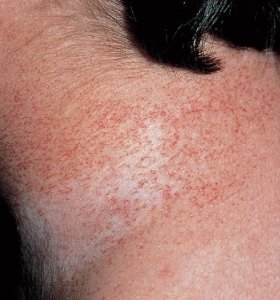
The patients will usually present with 0.5 to 2 cm multiple hypopigmented patches or thin plaques on the cheek and chin. The patches could be oval, round, or irregular in shape with poorly defined edges. Usually, the patients will have absent or minimal itch. The dryness and scaling are more noticeable in the winter, while the hypopigmentation is more noticeable in summer.

Pityriasis alba patches undergo several stages:

  1. The appearance of scaly pink with just a palpable surface.
  2. The hypopigmentation stage with fine surface scale.
  3. The post-inflammatory hypopigmented macule/ patch with no scale.
  4. Resolution of the lesion. 

Diagnosis:

The diagnosis is usually clinical, and the investigations are done to rule out other differential diagnosis:

  • Wood lamp: No enhancement in the cases of pityriasis alba.
  • Scrapings for mycology: negative in the cases of pityriasis alba.
  • Skin biopsy: Usually not indicated, would reveal spongiotic dermatitis and melanin reduction.

A dermoscopy of pityriasis alba will reveal, a poorly defined pale area, white structureless areas, superficial white scale, and normal hair color. 

Management:

Asymptomatic pityriasis alba requires no treatment. A moisturizing cream to improve the dryness, and a mild topical steroids will reduce redness and itchiness if present. It has been observed that calcineurin inhibitors, such as tacrolimus ointment and pimecrolimus cream, speed up the return of skin color and maybe just as effective as hydrocortisone.

Psoralen + UVA photochemotherapy is used in severe situations, this treatment may aid in repigmentation; nevertheless, the likelihood of recurrence following treatment discontinuation is considerable. Laser therapy can be effectively treated with a twice-weekly, 308-nm excimer laser treatment for 12 weeks. 

Prevention:  

Sunscreen use could be utilized to reduce the development of pityriasis alba.

Outcome: 

Pityriasis alba usually goes away in a year, although it might take anything from a few months to two or three years. The color gradually reverts to its original state.

Written by:

Mohammed Alahmadi, Medical Student. 

Revised by:

Maee Barakeh, Medical Student. 

References:

DermNet.

Fitzpatrick’s Color Atlas and Synopsis of Clinical Dermatology.

Dermatology by Bolognia.

Erythema Dyschromicum Perstans ( Achy Dermatosis )

Definition:

It is a type of acquired macular hyperpigmentation that occurs on the face, neck, and trunk. It is characterized by distinct circular or irregular patches that have a gray color. Erythema dyschromicum perstans is also known as ashy dermatosis (of Ramirez) due to its color. 

Epidemiology:

The disease typically manifests in the second or third decade of life, with no predilection for a particular gender. Lesions, however, can develop in old people or very young children.

Etiology:

The precise etiology of erythema dyschromicum perstans remains unknown. Due to its histological characteristics, it is frequently categorized as a form of lichen planus pigmentosus. There are some theories including:

-Genetic predisposition

-Allergic reaction to cosmetics and hair dye

-Viral infections 

-An adverse effect of drugs and medications

Clinical features:

-The pigmented patches might be either symmetrical or unilateral in their distribution.

-Early lesions may have an elevated appearance, a more distinct border, and a reddish color. This stage is not necessarily noticeable.

-Other than that, the patient is well and has no related illnesses or abnormal blood tests.

Diagnosis:

Occasionally, the clinical presentation of erythema dyschromicum perstans may exhibit sufficient characteristic features to provide a diagnosis of the condition. A skin biopsy can show mild vacuolar degeneration of the basal layer in initial lesions, and pigmentary incontinence with dermal melanophages in more developed patches.

Management:

Erythema dyschromicum perstans shows a high level of resistance to the treatments currently available. Although it may remain unchanged for years, some cases ultimately resolve on their own.

Possible interventions to enhance appearance of erythema dyschromicum perstans include the following: 

-Topical corticosteroids

-Ultraviolet radiation exposure 

-Pigment lasers, such as the Q-switched ruby laser

-Chemical peels

Clofazimine has proven to be the most effective systemic therapy. Dapsone, griseofulvin, hydroxychloroquine, isoniazid, and corticosteroids have demonstrated efficacy in a few cases.


Written by:

Mashael Alanazi, medical student.

Resources:

DernNet.
Bolognia Textbook of Dermatology.

Image resource: Bolognia Textbook of Dermatology.

Tinea Versicolor

What is Tinea Versicolor?

Tinea versicolor is a commonly occurring, benign, superficial cutaneous fungal infection that is typically characterized by patches and macules on the back and chest that are either hyper- or hypopigmented. It is caused by Malassezia species. Despite the availability of efficient oral and topical treatment methods, disease recurrence is frequent.


Epidemiology of Tinea Versicolor

Tinea versicolor occurs worldwide, with a higher occurrence in tropical climates. Adolescents and young adults are the most common age groups to develop tinea versicolor. It can also impact children and infants.


Risk Factors for Tinea Versicolor

There are various risk factors associated with this disease. These risk factors include exposure to hot and humid weather, hyperhidrosis, use of topical skin oils, genetic predisposition, immunosuppression state, malnutrition, corticosteroid usage, and Cushing disease. It may clear in the winter and reoccur each summer.


Cause of Tinea Versicolor

Tinea versicolor does not result from a dermatophyte infection. It is considered non-contagious. The causative organism is a Malassezia species. It is a component of the normal skin flora, but it can cause the disease when it transforms into its pathogenic hyphal form. The reasons for the transformation of tinea versicolor are likely to be multifactorial.


Clinical Features of Tinea Versicolor

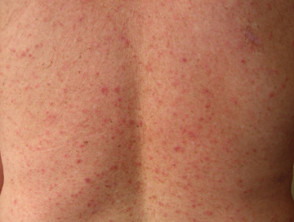
Tinea versicolor is typically asymptomatic; some complain of mild pruritus, with the main issue being its appearance.Tinea versicolor lesions are characterized by hyperpigmented, hypopigmented, or erythematous patches, macules, or thin plaques with a mild scale. To demonstrate this scale, the skin surface may need to be scratched. These lesions are usually small (<15 mm) and range in color from coppery brown, paler than the surrounding skin, or pink. The most typical areas of involvement are the upper trunk and proximal upper extremities. Less frequently, faces (common in children) or intertriginous areas are affected.


Diagnosis of Tinea Versicolor

Tinea versicolor is usually diagnosed clinically. Dermatoscopy and Wood’s lamp examination may show signs suggestive of tinea versicolor but do not confirm the diagnosis. Dermoscopic examination reveals fine-scale, nonuniform pigmentation within lesions and perilesional hyperpigmentation or hypopigmentation. Wood’s lamp examination exhibits yellow to yellow-green fluorescence in certain patients. The KOH preparation findings in tinea versicolor are considered diagnostic. It shows hyphae and yeast cells that resemble spaghetti and meatballs.


Management of Tinea Versicolor

The management of Tinea versicolor can be summarized into a few fundamental aspects:

1:Patient education

2: Topical antifungals, which are considered first-line therapy, including Selenium sulfide, zinc pyrithione, azoles (miconazole or ketoconazole), or allylamines (terbinafine)

3: Oral antifungals are only for infections that are severe, extensive, or resistant to topical treatment (not commonly used in children). Oral itraconazole and fluconazole are the recommended oral medications.


Recurrence of Tinea Versicolor

Tinea versicolor has a high recurrence rate. Preventive treatment with topical or oral medication administered intermittently is required to avoid recurrences in most cases.


Written by:

Atheer Alhuthaili , medical student. 

Revised by:

Maee Barakeh, medical student.

References:

Bolognia textbook of dermatology.

DermNet.

UpToDate.

Medscape .

Harlequin Ichthyosis 

A severe genetic variant of ichthyosis called harlequin ichthyosis manifests at birth as thicker, rigid skin plates that cover the entire body like armor. The condition is uncommon and equally affects men and women. Approximately one in 300,000 neonates are affected by it. Harlequin ichthyosis is not known to be racially predisposed. There could be a higher occurrence in societies where paternal consanguinity is prevalent.


Causes:

Harlequin ichthyosis is inherited in an autosomal recessive manner. The ABCA 12 gene alterations are to blame. It is thought that the ABCA12 gene encodes a transporter protein that is necessary for healthy skin development and is involved in the movement of epidermal lipids across cell membranes. The cell cannot produce any ABCA12 protein due to certain mutations in the ABCA12 gene. Additional mutations result in the synthesis of an atypically tiny protein that is incapable of carrying lipids in an appropriate manner. Harlequin ichthyosis is characterized by hard, thick scales that are caused by a disruption in the normal development of the epidermis caused by a loss of functional ABCA12 protein.


Clinical features:

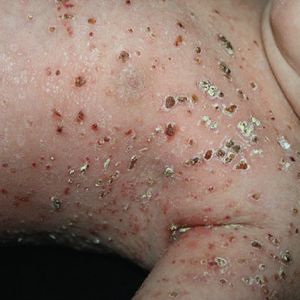
The clinical features include skin changes present at birth, significantly thickened, with deep erythematous fissures separating the scales and huge, glossy plates of hyperkeratotic scale. Facial features include degraded nasal alae and blocked nares. The external auditory canal may be blocked by scale, and the ears’ pinnae and retroauricular folds may be tiny or nonexistent. The lips’ traction results in eclabium.


Diagnosis:

Genetic laboratory testing and a physical examination of the patient are necessary for the diagnosis of harlequin ichthyosis. The most precise diagnostic procedure for harlequin ichthyosis involves looking for a loss of function mutation in the ABCA12 gene using genetic testing. Gene mutations may result in smaller versions of the proteins essential for skin development as well as poor lipid transport in the skin. A collodion membrane and congenital ichthyosiform erythroderma-like appearance are the outcomes of less severe mutations. Histology of the skin biopsy reveals hypergranulosis, parakeratosis, and a significantly thicker stratum corneum.


Management:

Treatment for harlequin ichthyosis focuses on skin protection and infection control; there is no known cure. For several weeks during their early years, the majority of harlequin babies require one-on-one nursing care. Infection prevention or treatment during this period may also require antibiotics. After bathing, when the skin is still damp, softening emollients especially those that contain urea, salicylic acid, or alpha hydroxy acids work exceptionally well. These products help to maintain the skin’s suppleness and moisture content while guarding against the fissures and cracks that can result in a subsequent bacterial infection. It has also been demonstrated that early systemic treatment with oral retinoids (such as acitretin or isotretinoin) improves overall survival, softens, or resolves plate-like scales, and heals skin fissures.


Complications: 

Poor immunity, infections, muscle shortening, limb necrosis and amputation, and respiratory failure.


Outcome:  

Infants with harlequin ichthyosis in the past hardly made it past the first few days of life. The survival rate has increased due to innovations in newborn care; globally, rates are getting close to 50%.  Youngsters who make it through the neonatal stage typically acquire a less severe form of ichthyosis, but they still exhibit fish-scale scales, seborrheic patches that retain waxy, yellowish material, generalized poor hair growth, and scarring alopecia.  Those who survive harlequin ichthyosis may nevertheless experience hypothyroidism, small stature, arthralgias, digit contractures, and failure to flourish.   


Written by:

Mohammed Alahmadi, Medical Student. 

Revised by:

Maee Barakeh, Medical Student. 

References:

DermNet

Dermatology by Bolognia

Fitzpatrick’s Color Atlas and Synopsis of Clinical Dermatology

Image source: Fitzpatrick’s Color Atlas and Synopsis of Clinical Dermatology 


ORF

Orf is a skin infection caused by parapox virus, a zoonotic contracted from goats and sheep. Because the condition causes crusts and scabs around the mouth, nose, teats, and other areas, it is also known as contagious pustular dermatitis. The disease lasts for roughly six weeks, and it is extremely contagious.


Cause and Mode of Transmission: 

Direct inoculation of contaminated material, frequently through an already-existing wound or scratch, results in lesions in humans. Shearers, freezer workers, sheep producers, and anyone bottle-feeding a lamb run the danger of contracting orf. Youngsters may potentially get it from playing in a sheep shed or on contaminated pasture. There have been reports of human orf following unintentional contact with animal-targeted orf virus vaccination. It is not typically spread from person to person, however sporadic reports of this have occurred.


Clinical Features:

The incubation period lasts from 5-6 days. Patients usually present with a 2-3 cm solitary small, firm, red lump that enlarges to form a flat-topped, blood-tinged pustule or blister on the forearms, hands, and fingers. Large lesions could be observed in immunocompromised individuals. 

Typically, making an incision through the skin will reveal firm, red tissue beneath, despite the appearance of pus from the skin. In the early stages, the orf lesion is frequently sensitive and irritating. Lymphangitis and lymphadenopathy of the inner side of the elbow and/or under the arm are not uncommon. These features could be associated with mild fever.  


Diagnosis:

A clinical diagnosis of orf is typically made in a person who has experience working with sheep or goats. PCR can be used to confirm the infection in vesicular fluid, skin biopsies, or viral swabs. Electron microscopy can be used to identify it as well. 


Management:

Most of the time, no special care is required because orf resolves on its own in roughly six weeks. While it is extremely rare, the lesion should be covered to avoid infecting the surrounding area or other persons. Shave excision is one method of removing large lesions. Antibiotics are the appropriate treatment for secondary bacterial infections. A few cases of orf have been found to respond well to imiquimod cream.


Complications: 

The complications of orf include secondary bacterial infection, erythema multiforme, toxic eythemas, and rarely pemphigoid.


Prevention:  

Farmers who raise sheep and goats should be mindful of the risk of orf and handle the animals especially lambs and young while using non-porous rubber gloves with washing hands. It is advised to vaccinate the herd using a live virus in the event of a severe orf outbreak.


Written by:

Mohammed Alahmadi, Medical Student. 

Revised by:

Maee Barakeh, Medical Student. 

References:

DermNet.

Fitzpatrick’s Color Atlas and Synopsis of Clinical Dermatology.

Andrews’ Diseases of the Skin: Clinical Dermatology.

Nodular Prurigo

Nodular prurigo (NP), also known as prurigo nodularis, is a chronic dermatological disorder characterized by intensely pruritic, firm nodules. Although it can form anywhere on the body, a symmetrical distribution of arms, legs, back, and torso is where it most frequently appears.


Etiology:

It is still unclear what specifically causes NP. Based on skin biopsies, it is believed to be related to a dysregulation of the nerves, with more nerve cells seen in the dermis and fewer in the epidermis. 

There is also an association with atopy, certain drugs, and systemic conditions such as chronic kidney disease, liver disease, diabetes, and malignancy.


Clinical features:

The severity of NP can range from a few to hundreds of lesions of different sizes.

At first, the rash:

– Include tiny, red, or pink bumps typically 3–8 mm in diameter.

– Has a hyperpigmented border

– Very itchy 

– Is distributed in readily accessible body regions, including the lateral aspects of the arms and legs, shoulders, chest, and buttocks.

Sometimes, it begins as a papule and progresses to a nodule or plaque.

Scratching results in:

-Enlargement producing a raised, warty, nodular surface. The nodules have a symmetrical distribution and are usually hard and small, but they can reach up to 3 cm in diameter.

-Ulceration of the skin that can lead to an increased risk of infection.

-Healed lesions may leave a scar or discoloration.


Diagnosis:

The diagnosis of NP is clinical, based upon the clinical presentation of distinctive excoriated, nodular lesions frequently distributed symmetrically and a patient’s history of persistent, intense pruritus. A skin biopsy is not routinely performed to confirm the diagnosis.

It is essential to evaluate systemic causes of persistent pruritus in individuals with NP who do not have a history of atopic dermatitis or other pruritic skin disorders. These causes may include chronic kidney disease, liver disease, thyroid disease, HIV infection, parasite infestation, or malignancy. The initial laboratory and imaging evaluation may consist of:

  • Complete blood cell count
  • Liver function tests
  • Blood urea nitrogen and creatinine
  • Thyroid-stimulating hormone
  • HIV test
  • Urinalysis
  • Stool examination for ova and parasites
  • Chest radiograph


Management:

A complex and multi-modal strategy is necessary for the treatment of NP which includes:

  • Instructing patients on various ways to reduce skin itching and scratching 
  • Treating pruritus symptoms 
  • Topical or systemic therapies that target the itch-scratch cycle and minimize skin lesions

Common treatment options include: 

  • Topical emollients
  • Topical steroid cream or ointment — usually ultrapotent steroid and applied under hydrocolloid dressing or paste bandage occlusion, or steroid impregnated tape
  • Topical capsaicin and tacrolimus 0.1% ointment for itch
  • Local corticosteroid injection into the nodules to reduce inflammation
  • Sedating oral antihistamines
  • Phototherapy with either UVB or photochemotherapy (PUVA)
  • Systemic treatment with gabapentin, pregabalin, and naltrexone
  • Occasionally, antibiotics may be used to treat locally infected skin (cellulitis).


Written by:

Mashael Alanazi, Medical student.

Revised by:

Maee Barakeh, Medical student.

References:

DermNet

Uptodate

Molluscum Contagiosum

It is a benign, often asymptomatic viral infection of the skin, with no systemic manifestation. It is caused by molluscipoxvirus, a part of the poxviridae family which is a family of double-stranded DNA viruses. It is most common in childhood and early adolescence. 


Risk Factors:

As mentioned, the etiology is due to molluscipoxvirus, but we have several risk factors associated with this disease. These risk factors include immunosuppression state, active atopic dermatitis, hot and humid climates, and crowded living conditions. Generally, those with poor immune function and immunocompromised skin are at risk for molluscum contagiosum.


Mode of Transmission:

Molluscum contagiosum can be transmitted due to direct contact during sports or other activities including sexual contact, indirect skin contact, or with fomites which are any object that can transmit the organism. Lastly, auto-inoculation can transmit the lesion by scratching or any trauma resulting in linear lesions (Pseudo koebner phenomenon).  


Clinical Features:

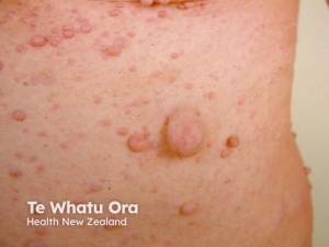
The incubation period will usually be 2-6 weeks or more. Usually, the patient will come complaining of single or multiple non-tenders, skin-colored, pearly, dome-shaped papules with central umbilication. The lesion will be 2–5 mm in diameter and contain a caseous plug which if you squeeze will produce white cheesy fluid that consists of molluscum bodies. 

The usual lesions in children will be the face, trunk, and limbs, in contrast to adults where the lesions will be usually in the abdomen, inner thighs, and genitalia. Finally, patients with HIV will usually have widespread and large (more than 15 mm) lesions. 


Diagnosis:

Molluscum contagiosum is usually recognized by its characteristic clinical appearance (dome-shaped papules, often with central umbilication), and is distinctive on dermatoscopy which will show white molluscum bodies can often be expressed from the center of the papules. Sometimes, the diagnosis is made on skin biopsy. Histopathology will show the following features, acanthosis, cup-shaped invagination, and molluscum bodies (Henderson-Paterson bodies) which are keratinocytes with eosinophilic intracytoplasmic inclusion bodies containing viral particles. 


Management:

Spontaneous remission is expected, however active treatment is considered in sexually transmitted molluscum, immunocompromised individuals or upon parental request.  

The first line of treatments includes cryotherapy and curettage which has an immediate resolution of the lesion. Both treatments  are well tolerated by adults.

Cantharidin which is a blistering agent can be applied directly to lesions, and a small blister will form which will result in the disappearance of the lesion and healing without scarring. Podophyllotoxin can also be used directly on the skin. 

Immunocompromised patients or individuals with refractory disease will usually benefit from Cidofovir, Interferon-α, and Imiquimod. Other treatments for molluscum include potassium hydroxide, salicylic acid, topical retinoids, oral cimetidine, and pulsed dye lasers.


Complications: 

It includes secondary bacterial infection from scratching, conjunctivitis, disseminated secondary eczema, and scarring.


Prevention: 

Patients should be instructed to do the following, keep good hygiene, cover the lesion, do not share towels, clothing, or other personal items, and avoid sexual activity until the resolution of the lesion.


Written by:

Mohammed Alahmadi, Medical Student. 

Revised by:

Maee Barakeh, Medical Student. 

References:

DermNet

Dermatology Essentials by Bolognia

Fitzpatrick’s Color Atlas and Synopsis of Clinical Dermatology

UpToDate  

Keratoacanthoma

Keratoacanthoma (KA) is a commonly encountered skin tumor that presents a captivating clinical challenge due to its rapid growth and locally destructive nature. While some cases of KA may regress spontaneously, they often leave behind visible scarring. However, the clinical presentation of KA can closely resemble well-differentiated squamous cell carcinoma (SCC), leading to unpredictability in its course.

Pathophysiology:

The precise pathophysiological mechanisms underlying keratoacanthoma are not fully understood. However, it is widely believed to arise from the hair follicles or the sebaceous glands. Several hypotheses have been proposed, including the role of ultraviolet radiation, genetic factors, viral infections, and immune dysregulation in the development of keratoacanthoma.

Clinical Features:

Keratoacanthoma typically presents as a rapidly growing dome-shaped nodule with a central crater filled with keratin debris. It commonly occurs on sun-exposed areas of the body, such as the face, scalp, ears, and dorsum of the hands. Initially, the lesion may resemble a pimple or a wart, but it rapidly enlarges over a period of weeks to months. The growth phase is followed by a stabilization phase, during which the lesion may persist for several months before spontaneously regressing.

Diagnosis:

The diagnosis of keratoacanthoma is primarily based on clinical examination and histopathological evaluation. A thorough physical examination, including a dermatoscopic assessment, can aid in distinguishing keratoacanthoma from other skin lesions. Dermoscopy often reveals a central plug, keratin-filled crater, and peripheral keratotic rim. To confirm the diagnosis, a skin biopsy is performed, and the specimen is sent for histopathological analysis. Microscopically, keratoacanthoma is characterized by a well-demarcated cup-shaped lesion with a central keratin-filled crater, hyperkeratosis, and proliferation of keratinocytes.

Management:

The management of keratoacanthoma depends on various factors, including the size, location, and individual patient characteristics. Several treatment options are available, and the choice of therapy should be tailored to the specific case. Here are some commonly employed management strategies:

1. Observation: In some cases, keratoacanthomas may undergo spontaneous regression without any intervention. Close observation with regular follow-up visits may be appropriate for small, asymptomatic lesions in low-risk areas.

2. Surgical Excision: Surgical excision is a commonly employed treatment modality for keratoacanthoma. It involves complete removal of the lesion with a margin of healthy tissue to reduce the risk of recurrence. This approach is particularly suitable for larger or symptomatic tumors.

3. Mohs Micrographic Surgery: Mohs surgery is a specialized technique that ensures the highest cure rate while preserving as much healthy tissue as possible. It is often recommended for lesions in cosmetically sensitive areas or those with aggressive features.

4. Topical Therapies: In select cases, topical treatments such as imiquimod cream or 5-fluorouracil may be employed. These medications stimulate the immune system or inhibit cell proliferation, leading to tumor regression.

5. Cryotherapy: Cryotherapy involves freezing the lesion with liquid nitrogen. It is commonly utilized for smaller keratoacanthomas and can be performed in an outpatient setting.

Written by:

Deemah AlHuraish, medical student.

Revised by:

Maee Barakeh, medical student.

References:
DermNet

NCBI

AOCD

Cold Urticaria

Cold urticaria is one type of chronic inducible urticaria that is relatively uncommon. Exposure to cold air, water, or objects cause the development of urticarial weals.

Epidemiology:

About 0.05% of the population develops cold-induced urticaria over time. Though often diagnosed in people of young and middle-age, it may also manifest in children and the elderly. It is twice as common in females as in males.

Etiology: 

Cold urticaria is a condition that arises as a result of exposure to low temperatures. The mechanism by which the cold stimulus induces the activation of mast cells and the subsequent release of histamine, along with other inflammatory mediators, remains unclear.

Cold urticaria can manifest as either primary (idiopathic) or due to an underlying hematologic or infectious illness. The majority of cases fall under the category of idiopathic, meaning that the cause is unknown.

Clinical features:

The symptoms of cold urticaria manifest within a time frame of 2 to 5 minutes following exposure and often persist for a duration of 1 to 2 hours.

Urticaria and angioedema can manifest as either localized, affecting specific areas of the body, or generalized, resulting in a rash that covers the entire body.

Systemic reactions, from mild discomfort to anaphylaxis, have been linked to prolonged exposure to cold, such as when swimming in cold water. Cold-water aquatic activities are dangerous because of the risk of anaphylaxis and secondary drowning.

Patients undergoing surgery under general anesthesia or in cold rooms are also at risk for developing severe reactions.

Diagnosis:

The diagnosis of cold urticaria may be established with the application of an ice cube to the skin of the forearm for a duration of 1 to 5 minutes. If the patient has cold urticaria, it is expected that a clearly discernible red and swollen rash would develop within a few minutes in the region that has been subjected to the cold-stimulation test.

Associated illnesses may also be identified by doing complete blood count and metabolic testing.

Treatment:

It is important for patients with cold urticaria to take precautions against experiencing a sudden decrease in body temperature. Supervision is required at all times during any aquatic activity (including swimming and surfing).

Patients at risk for anaphylaxis should always have access to adrenaline.

Non-sedating antihistamines at large dosages (up to 40 mg of cetirizine daily, for instance) may be useful. A brief course of oral glucocorticoids may help patients with severe angioedema.

Cautious induction of cold tolerance may be effective (desensitization) by progressively hardening the skin to cold temperatures and then exposing the skin to it frequently, for example, by taking regular cold showers.

Written by:

Mashael Alanazi, medical student

Revised by:

Maee Barakeh, medical student

Resources:

DermNet

UpToDate

Merkel Cell Carcinoma

Merkel Cell Carcinoma, also known as cutaneous neuroendocrine carcinoma, is a rare and aggressive form of skin cancer with a high risk for recurring and metastasizing. It occurs almost exclusively in white skin, and is slightly more common in males.

Etiology:

Approximately 80% of examined Merkel cell carcinomas have Merkel cell polyomavirus (MCPyV) identified. It is believed that when immune function is compromised, the virus induces gene alterations that result in Merkel cell cancer.

Because virus-negative tumors typically develop on skin that has been exposed to the sun, they are linked to high UV radiation exposure. One major contributing factor to the development of Merkel cell carcinomas is immunosuppression.

It was once thought that Merkel cells, which are skin pressure receptors, were the source of Merkel cell cancer. Based on clonal immunoglobulin chain rearrangement, early B-cell markers expressed, and cellular shape, a new study suggests that their ancestors were lymphocytes.  

Clinical features:

Typically, asymptomatic, non-tender, solitary, uneven red nodule that is rapidly growing is the first sign of Merkel cell cancer. Although it grows far more quickly, it frequently resembles other, more prevalent skin malignancies in appearance, such as basal cell carcinoma.

Multiple metastases may form surrounding the primary tumor in Merkel cell tumors, which spread through the lymphatic system and can cause a local recurrence. Additionally, it may spread to axillae, groin, and neck lymph nodes.

The rate of recurrence  is markedly higher than that for invasive melanoma, squamous cell carcinoma, or basal cell carcinoma. The majority of recurrences happen in the first two years following diagnosis.

Diagnosis:

Any tumor exhibiting the following clinical characteristics, which are seen in roughly 90% of patients, should be ruled out as being likely to be Merkel cell carcinoma. These characteristics can be remembered using the acronym “AEIOU”:

  • Asymptomatic or non-tender
  • Expanding rapidly
  • Immune suppressed
  • Older than 50
  • UV-exposed fair skin

A tumor biopsy is the primary test. This demonstrates the typical histology of Merkel cell cancer. Since thyroid transcription factor (TTF1) is typically negative and cytokeratin-20 (CK20) is positive in up to 95% of tumors, immunohistochemistry can be useful.

If the tumor has progressed to other places, a general examination, including assessment of the local lymph nodes, and staging studies may be scheduled. Imaging modalities, lymph mode ultrasound scanning, and sentinel node biopsies are examples of staging investigations. 

Management:

Treatment options include excision or Mohs surgery.  Except in very small lesions, radiation therapy is usually administered to the Merkel cell carcinoma site along with regional lymph node placement.

Pembrolizumab and nivolumab, immune checkpoint inhibitors for advanced Merkel cell carcinoma, up to 50% of patients have shown positive response. Avelumab, another PD-1/PD-L1 medication, received expedited approval from the US Food and Drug Administration in March 2017 to treat metastatic Merkel cell carcinoma.

Prognosis: 

Five-year survival rates are overall 30–50%. Survival rates are better for patients initially diagnosed with Stage 1 Merkel cell carcinoma and in younger patients than when it has already metastasized or patients are over 80 years of age. 

Written by:

Mohammed Alahmadi, Medical Student. 

Revised by:

Maee Barakeh, Medical Student. 

References:

DermNet

Fitzpatrick’s Color Atlas and Synopsis of Clinical Dermatology

Review of Dermatology by Ali Alikhan

Hand, Foot, and Mouth Disease

Hand, foot, and mouth disease (HFMD, also called enteroviral vesicular stomatitis is a common acute viral infection, primarily affecting young children under the age of 5. It is known to appear in a characteristic distribution, involving mucocutaneous lesions over hands, feet and oral cavity.

Etiology:

HFMD transmission is very rapid, mainly via  direct contact with the blister fluid or droplets spread from the mouth.

Numerous coxsackie virus serotypes have been implicated to cause HFMD, most commonly type A16.  Enterovirus 71 infection has been associated with cardiopulmonary and neurologic complications and even deaths in affected young children.


Clinical Features:

Patients with HFMD often present with fever and malaise, and they may have a mild prodrome prior to the onset of the exanthem.

HFMD exanthem is characterized by:

  •  vesicular eruption on the palms and soles. The dorsal aspect of the hands and feet may be involved as well as the buttocks and perineum. The lesions are typically tender and are usually oval rather than round.
  • Oral lesions are most common on the tongue, buccal mucosa, palate, uvula, and anterior tonsillar pillars. They are usually painful, especially with oral intake.
  • Onychomadesis occasionally occurs months after HFMD as a consequence of temporary nail matrix arrest related to the viral infection.


Complications: 

HFMD rarely causes serious complications. Enterovirus 71 is usually associated with more severe infections and complications, especially among immunosuppressed individuals or neonates. Rare serious reported complications include myocarditis, meningoencephalitis, and pulmonary edema.


Diagnosis:

HFMD is diagnosed clinically based on exposure history, geographic location, and clinical signs and symptoms.

Confirmation of an enteroviral infection may be accomplished by viral testing from vesicular fluid via reverse transcription polymerase chain reaction (RT-PCR)-based assays to determine  the specific type of enterovirus, however, it is done only for research and public health purposes. 


Management:

HFMD is a self-limiting disease  with spontaneous resolution typically within 1–2 weeks. Supportive care is the mainstay of treatment, including hydration, analgesic and blister care.

Pleconaril has been demonstrated in recent research and has shown a promising outcome for serious enteroviral infections. It is a specific antiviral therapy that interferes with enterovirus attachment and uncoating by binding to the protein capsid.


Written by:

Maee Barakeh, medical student.

References:

Bolognia textbook of dermatology

DermNet